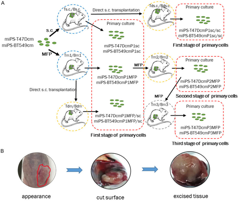
Figure 1.

A. Schematic drawing of the preparation of primary cells. The first stage: miPS-T47DcmP1sc, miPS-BT549cmP1sc, miPS-T47DcmP1sc/sc, miPS-BT549cmP1sc/sc, miPS-T47DcmP1MFP, miPS-BT549cmP1MFP, miPS-T47DcmP1MFP/sc and miPS-BT549cmP1MFP/sc cells from the tumors developed from the transplantation of miPS-T47Dcm and miPS-BT549cm cells in BALB/c-nu/nu mice. The second stage of primary cells is the P culture of the Tm2/Bm2 tumors. The third stage of primary cells is the P culture of the Tm3/Bm3 tumors. Each stage of primary cells is surrounded by red broken squares. Ts.c./Bs.c. and Tm1/Bm1 are the primary tumors (blue broken circles). Tds.c./Bds.c., Tm2/Bm2 and Tdm/Bdm are the secondary tumors (yellow broken circles). Tm3/Bm3 is the tertiary tumor (gray broken circles). B. The macroscopic features of the Tm1 tumor transplanted into the MFPs. The border of the tumor was ill defined (red line) with a firm consistency that was too hard (left). The cut surface shows hemorrhage and necrotic areas (middle). The base appears indurated and infiltrating (right).
