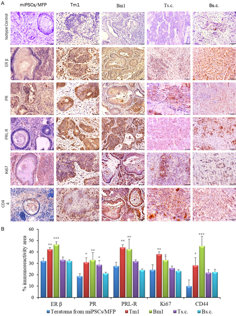
Figure 4.

IHC analysis of primary Tm1/Bm1 and Ts.c./Bs.c. tumors transplanted into the MFPs and s.c. tissue, respectively. A. Immunoreactivity to ERβ, PR, PRL-R, Ki67 and CD44 is shown in paraffin sections of Tm/Bm1 and Ts.c./Bs.c. tumors derived from miPS-T47Dcm and miPS-BT549cm cells. Scale bar = 64 µm at 20× magnification. B. Percentage of the immunoreactive area (mean ± SD) of ERβ, PR, PRL-R, Ki67 and CD44 in the different experimental groups. Each bar in the graphs represents the average of six readings (mean ± SD). Significance compared with tumors derived from miPSCs transplanted into the MFPs. *P<0.05, **P<0.01, ***P<0.001.
