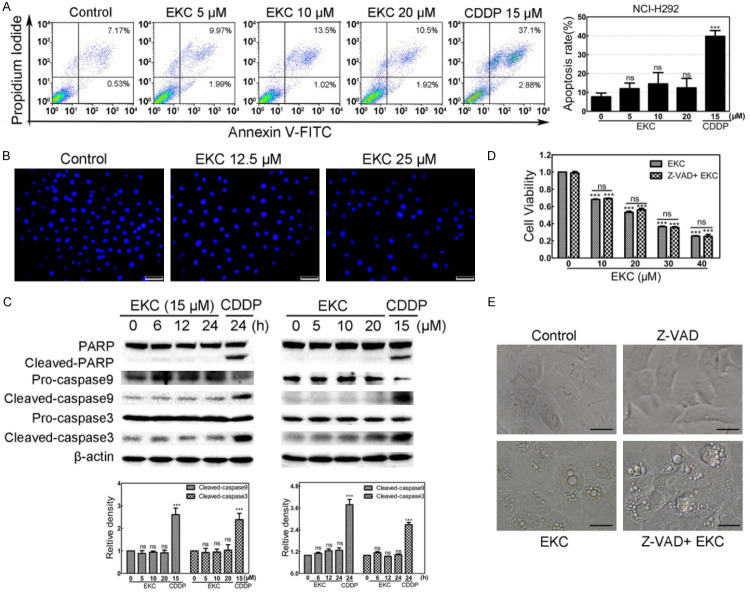
Figure 2.
EKC caused NCI-H292 cell death was distinct from apoptosis. A. NCI-H292 cells were treated with EKC at indicated concentrations for 48 h, stained with Annexin V/PI and followed by flow cytometry assay. Cisplatin (CDDP) 15 μM treatment was used as positive control. Data are expressed as mean ± SD, n = 3. B. NCI-H292 cells were treated with EKC at 12.5 and 25 μM for 24 h, the changes in nuclear morphology were evaluated by fluorescence microscopy after DAPI staining. Bar = 50 μM. C. NCI-H292 cells were incubated with EKC as indicated, cell lysates were subjected to immunoblotting for PARP, caspase9 and caspase3, β-actin was used as loading control. The band density was normalized to untreated group. D. Cell viability of NCI-H292 cells with or without Z-VAD (OMe)-FMK. Cells were pretreated with 20 μM Z-VAD (OMe)-FMK for 1 h before EKC treatment, cell viability was determined by MTT assay. Results were from thee separate experiments. Values are expressed as mean ± SD, n = 3. ***P < 0.001. E. NCI-H292 cells were pretreated for 1 h in the presence or absence of Z-VAD (OMe)-FMK (20 μM) prior to addition of EKC or DMSO. Phase-contrast images were captured after 12 h. Bar = 20 μM.

