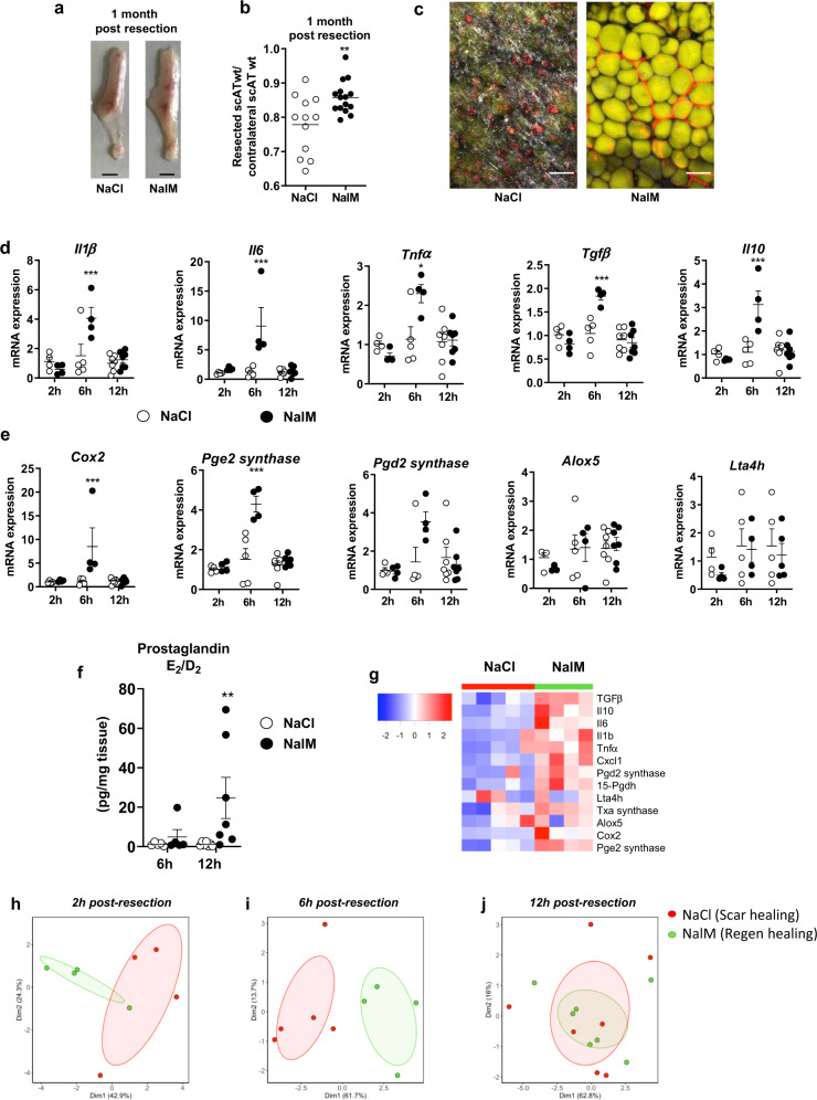
Fig. 1. Regenerative healing is characterized by early and transient inflammation.
a Representative pictures of scAT 1 month post-resection and NaCl (scar healing condition) or NalM (regenerative condition) treatment (scale bar: 0.5 cm). b Weight ratio between resected and contralateral scAT 1 month post-resection in NaCl or NalM-treated mice (n = 12–15 per group). c Representative pictures of resection plane area in NaCl and NalM treated mice one month post-resection. Adipocytes (yellow) were stained with Bodipy and vessels (red) with in vivo Lectin injection. Collagen fibers (gray) were imaged using a second harmonic generation (SHG) signal. Images were obtained using maximum intensity projections of 23 stack images. (scale bar: 50 µm). d, e Quantification by RTqPCR at 2 h, 6 h, and 12 h post-resection of mRNA encoding cytokines (Interleukin 1β, 6 (Il1β, Il6), Tumor Necrosis Factor α (Tnfα), Transforming Growth Factor β (Tgfβ) and interleukin 10 (Il10)) (d) and enzymes involved in lipid mediator synthesis (Cyclooxygenase 2 (cox2), Prostaglandin E2 synthase (Pge2 synthase), Prostaglandin D2 synthase (Pgd2 synthase), Arachidonate 5-lipoxygenase (Alox5) and Leukotriene A4 Hydrolase (Lta4h)) (e) in SVF isolated from the injured scAT of NaCl or NalM treated mice (n = 4–6 per group). f Quantification of PGD2 and PGE2 metabolites in the exudate of the resection plane of NaCl or NalM treated mice, 6 and 12 h post-resection. Results are expressed as a ratio between PGD2 and PGE2 metabolites (n = 5–7 per group). g Heatmap performed on 14 standardized gene expressions, 6 h post-resection. The dendrogram performed according to the Ward method was able to cluster between scar and regenerative healing conditions. Principal component analysis performed at 2 h (h) 6 h (i) and 12 h (j) post-resection on standardized gene expression. Individuals with scar healing and regenerative healing signatures were colored in red and green, respectively. Data are represented as mean ± SEM. (*p < 0.05, **p < 0.01, ***p < 0.001 between scar and regenerative healing conditions). NalM naloxone methiodide, scAT subcutaneous adipose tissue, SVF stromal vascular fraction, wt weight.

