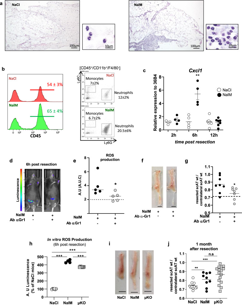
Fig. 2. Neutrophils depletion impairs ROS production and inhibits scAT regeneration in NalM-treated mice.
a Representative comparative microscopic aspects of the resection plane of scAT 8 h post-surgery in NaCl and NalM-treated mice (Hemalun & eosin staining). b Representative histograms and dot plot analyses of SVF cells isolated from NaCl (red curve) or NalM (green curve) treated mice 6 h post-resection, showing the percentage of CD45+ cells and granulocytes (neutrophils: CD45+/Ly6G+/Ly6C−/CD11b+ and monocytes CD45+/Ly6G−/Ly6C+/CD11b+) in the resection plane. c Gene expression quantification by RT-qPCR of the chemokine Cxcl1 in SVF isolated from the resection plane of NaCl-treated or NalM-treated mice 2, 6, and 12 h post-resection (n = 4 per group). d Representative in vivo imaging of ROS production 6 h post-resection in NalM-treated mice, treated or not with anti-Gr1 blocking antibody (Ab α-Gr1). e Quantification of ROS production in vivo from 0 to 72 h post-resection in NalM-treated mice, treated or not with Ab α-Gr1. The dotted lines represent ROS production obtained in NaCl-treated mice (n = 5 per group). f Representative pictures of scAT 1 month post-resection, in NalM-treated mice treated or not with Ab α-Gr1 (scale bar: 0.5 cm). g Weight ratio between resected and contralateral scAT 1 month post-resection in NalM treated mice, treated with isotype or Ab α-Gr1 (n = 7–8 per group). The dotted line show values obtained in scar healing (NaCl) conditions. h In vitro quantification of ROS production by Gr1+ populations sorted from scAT of NaCl, NalM, or µKO mice 6 h post-resection (n = 4-6 per group). i Representative pictures of scAT 1 month post-resection, in NaCl or NalM-treated mice and in NaCl-treated mice knock out for the µ opioid receptor (µKO) (scale bar: 0.5 cm). j Weight ratio between resected and contralateral scAT 1 month post-resection in NaCl, NalM-treated and µKO mice (n = 9–16 per group). Data are represented as mean ± SEM (*p < 0.05, ***p < 0.001 between scar and regenerative healing conditions). AT adipose tissue, AU arbitrary units, AUC area under the curve, NalM naloxone methiodide, scAT subcutaneous adipose tissue, SVF stromal vascular fraction, ROS reactive oxygen species, wt weight.

