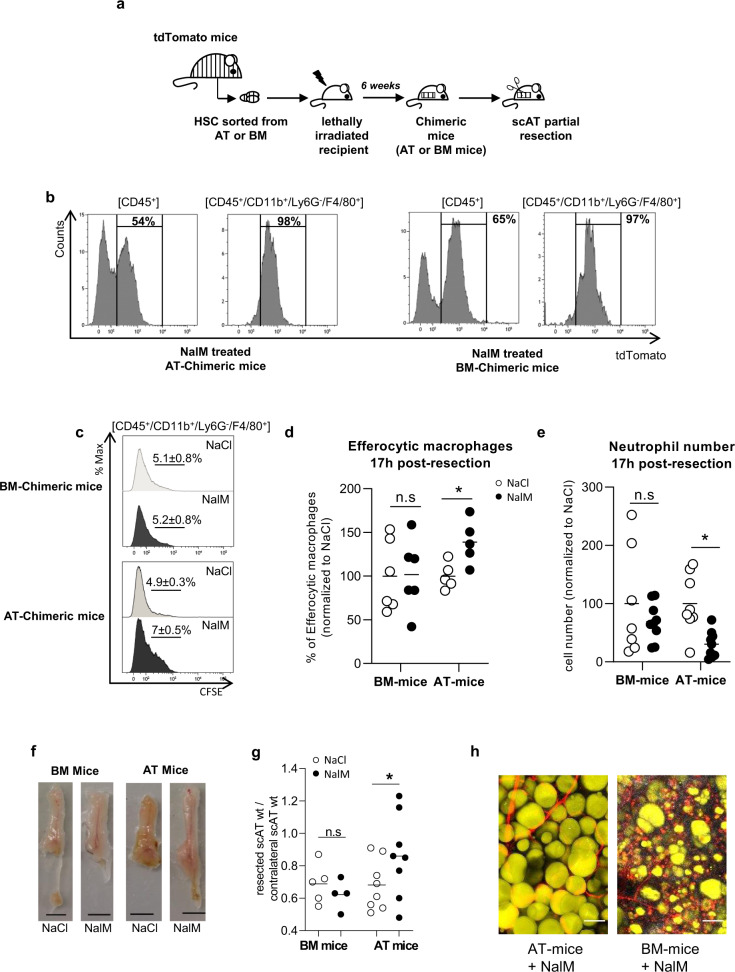
Fig. 4. AT Resident macrophages but not classical BM-derived macrophages are required for tissue regeneration.
a Hematopoietic chimera strategy: 2 × 10³ LSK cells sorted from the scAT or the bone marrow (BM) of mTmG mice were co-injected with 2 × 10⁵ total BM cells of C57Bl6 into lethally irradiated C57Bl6 recipients. Two months after hematopoietic reconstitution, scAT resection was performed and chimeric mice were treated with NaCl or NalM. b Representative histograms showing chimerism (tdTomato staining) in total immune cells (CD45+) and macrophages (CD45+/CD11b+/Ly6G-/F4/80+) in NalM-treated AT- and BM-chimeric mice, 24 h post-resection. c Representative histograms of CFSE staining in macrophages 24 h post-resection in NaCl and NalM-treated chimeric mice, corresponding to the percentage of macrophages having engulfed CFSE+ neutrophils. d Quantification of efferocytic macrophages 24 h post-resection in NaCl and NalM-treated chimeric mice (n = 5–6). e Quantification of neutrophil number by flow cytometry 24 h post-resection, in NaCl or NalM, treated mice (n = 7–8 per group). f Representative pictures of scAT 1 month post-resection, in BM- and AT-chimeric mice treated or not with NalM (scale bar: 0.5 cm). g Weight ratio between resected and contralateral scAT in AT- and BM-chimeric mice 1-month post-resection (n = 5–8 per group). h Representative pictures of resection plane area of NalM-treated AT- and BM- chimeric mice, one-month post resection. Adipocytes (yellow) were stained with Bodipy and vessels (red) with in vivo Lectin injection. Collagen fibers (gray) were imaged using second harmonic generation (SHG) signal. Images were obtained using maximum intensity projections of 23 stack images (scale bar: 50 µm). Data are represented as mean ± SEM. (*p < 0.05, **p < 0.01, ***p < 0.001 between scar and regenerative healing conditions). AT adipose tissue, BM bone marrow, LSK Lin−/Sca-1+/c−Kit+ cells, NalM naloxone methiodide, scAT subcutaneous adipose tissue, wt weight.

