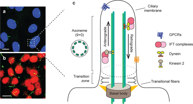
Fig. 1. Schematic structure of primary cilia.
Immunofluorescence images of primary cilia (green, ADCY3) in hypothalamic cells (a) and arcuate nuclei (b). Scale = 20 μm. Schematic structure of primary cilia (c). The primary cilium is an antenna-like organelle that receives diverse signals from the extracellular environment. It is comprised of the ciliary membrane surrounding the microtubule-based axoneme. The nine parallel microtubule doublets of the axoneme, which show “9 + 0” rings, form the backbone of the appendage, while the basal body acts as a microtubule-organizing center. The components that are transported from the basal body to the ciliary tip by anterograde transport rely on the intraflagellar transport (IFT) protein attached to the motor protein kinesin 2. In contrast, retrograde transport from the ciliary tip to the cytoplasm depends on dynein motor proteins. The ciliary membrane is highly enriched with several receptors, including G protein-coupled receptors (GPCRs).

