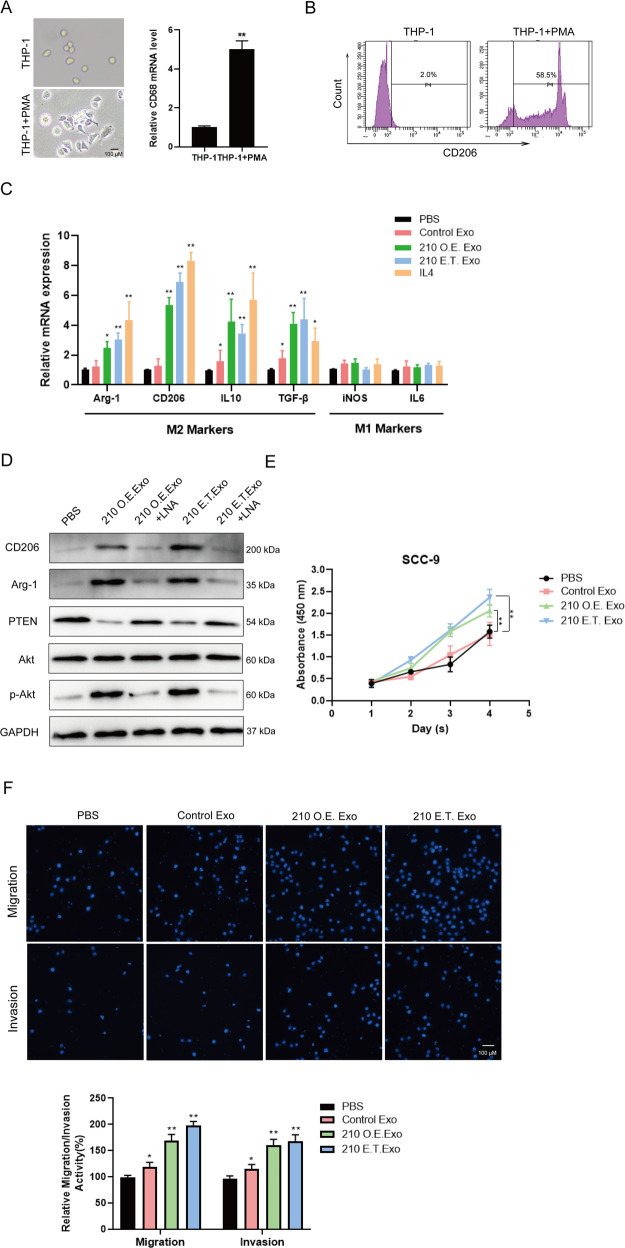
Fig. 7. Exosomal ANLN-210 promotes M2 polarization of macrophages.
A The representative cell morphologic photographs of macrophages. THP-1 monocytes were differentiated into macrophages using 150 nM Phorbol 12-myristate 13-acetate (PMA). The CD68 mRNA level was examined in THP-1 and PMA-induced macrophages (THP-1 + PMA). B Detection of macrophage marker CD206 by flow cytometry. C The relative expression of Arg-1, CD206, IL-10, TGF-β, iNOS, and IL-6 was measured using qRT-PCR in the following groups (PBS, Control Exo, 210 O.E. Exo, 210 E.T. Exo, and IL-4) “M2” macrophages markers: Arg-1, CD206, IL-10, and TGF-β. “M1” macrophages markers: iNOS and IL-6. *P < 0.05, **P < 0.01. D Western blot assays were performed to determine the protein levels of M2 markers CD206 and Arg-2, PTEN, p-Akt, Akt in macrophages treated with PBS, 210 O.E. Exo, 210 O.E. Exo + LNA, 210 E.T. Exo, 210 E.T. Exo + LNA. LNA: ANLN-210 locked nucleic acid. E Cell proliferation was analyzed in SCC-9 cells treated with macrophages incubated with PBS, Control Exo, 210 O.E. Exo, and 210 E.T. Exo. *P < 0.05, **P < 0.01. F Cell migration and invasion were analyzed in SCC-9 cells treated with macrophages incubated with PBS, Control Exo, 210 O.E. Exo, and 210 E.T. Exo. *P < 0.05, **P < 0.01.

