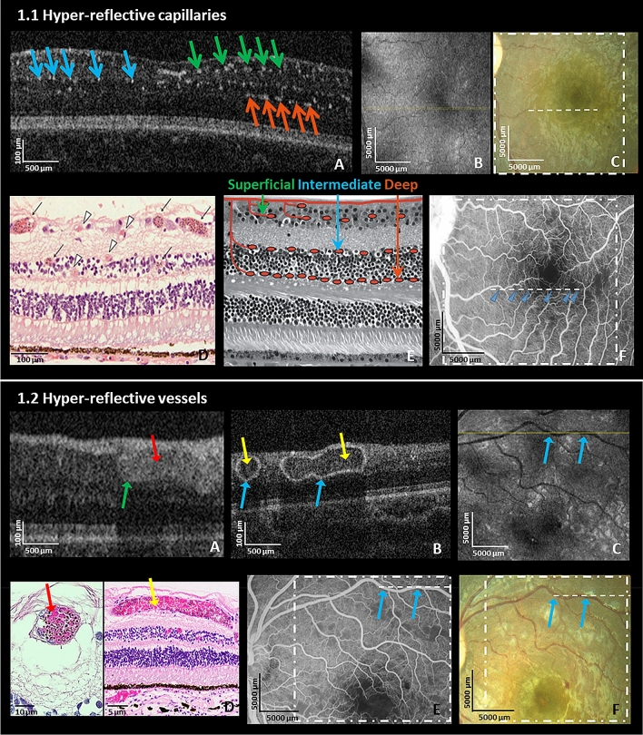
Figure 1.
Hyper-reflective capillaries and vessels. (1.1) Left eye of a 7-month old male CM patient on admission. (A) HH-OCT B-scan showing hyper-reflective dots corresponding to locations of capillaries (green, blue and orange arrows: superficial, intermediate and deep capillary plexus); (B) En-face OCT displays no visible change; (yellow line: location of the OCT B-scan in the image (A)). (C) Fundus photo; dashed line and square correspond to (A, B); (D) Representative retinal cross-section histology from a different fatal patient showing capillaries and venules affected by parasite sequestration (black arrows: intense sequestration; arrowheads: low or no sequestration); Reprinted with permission25; (E) Schematic representation of superficial, intermediate and deep capillary plexus collocated to capillaries filled with pRBCs on OCT in (A). Reprinted with permission24; (F) Fundus fluorescein angiography (FA) shows perfusion deficits of capillaries (blue arrows) corresponding to parasitized vessels on OCT in the same location of (1.1.A). (1.2) Left eye of a 42-month old male CM patient on admission. (A) Hyper-reflective vessel (blue arrow) corresponding to localization of vessel on fundus photo and FA (not shown) with hyper-reflective lumen (red arrow); and (B) Hyper-reflective vessels (blue arrows) with hypo-reflective lumina (yellow arrows) on OCT B-scans; (C) En-face OCT (yellow line: location of OCT B-scan in B, blue arrows: vessels shown in (B); (D) Representative retinal cross-section histology showing pRBCs in venules from a different fatal patient (red arrow shows vessels with pRBCs filling the lumen; yellow arrow shows a vessel where pRBCs cytoadhere to the vessel wall with red blood cells without parasites in the center of the vessel (similar to hypo-reflective lumina of blood vessels in (1.2.B); (E) Fundus fluorescein angiography and (F) Fundus photo corresponding to OCT in (1.2.B) (blue arrows corresponding to vessels in the image (B); dashed line and square correspond to (B, C).

