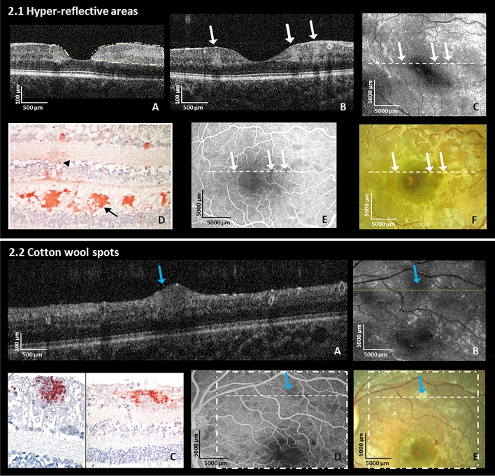
Figure 2.
Hyper-reflective areas and cotton wool spots (Retinal Whitening). (2.1.A, B) shows multiple hyper-reflective areas of various sizes (white arrows) located in the inner nuclear layer level, the outer plexiform layer and outer nuclear layer on OCT B-scans in the right eye of a 22-month old male CM patient and in the left eye of an 84-month old female CM patient at admission, respectively; (C) En-face OCT of the patient in (B); white arrows correspond to the location of white arrows in the image (B) (hyper-reflective areas); The hyper-reflective areas in OCT B-scan (B) show darkening on the en-face image (C) in this patient (white arrows); (D) Representative histology of immuno-histochemical staining for fibrinogen surrounding a small vessel (black arrowhead) mainly located in OPL (black arrow) from the literature (Reprinted with permission)17. (E) Fundus fluorescein angiography showing hypo-perfusion of capillaries and (F) fundus photo same eye as OCT in (B); white arrows and dashed lines show parafoveal whitening which are corresponding to the same fundus locations in (B, C, E, F). (2.2.A) Cotton wool spot (blue arrow) in the left eye at admission on OCT B-scan in the left eye of a 42-months old male CM patient at admission; (B) En-face OCT with darkening in area of cotton wool spot (blue arrow); fine yellow line: location of the B-scan in the image (A); blue arrow corresponds to blue arrow in image (A); (C) Immunohistochemical staining for b-APP in retinal nerve fiber layer (RNFL) from different CM patients. Reprinted with permission17. (D) Fundus fluorescein angiography showing grey area masking underlying capillaries (blue arrow) and (E) fundus photo from patient’s left eye corresponding to OCT showing white cotton wool spot (blue arrow) in (2.2.A). The dashed square and the dashed line correspond to en-face OCT in B and B-scan in (A). Blue arrows show the location of a cotton wool spot. Blue arrows correspond to the same fundus locations in (A, B, D, E).

