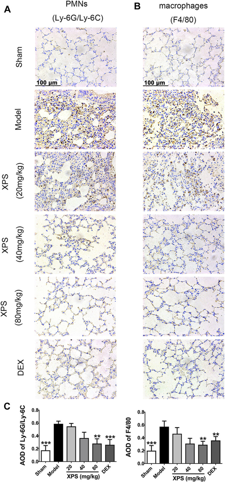
FIGURE 6.

XPS inhibited inflammatory cells infiltration in lung tissue of ALI mice. The PMNs (A) and macrophages’ (B) infiltration were observed under light microscopy (400×) by immunohistochemistry analysis. A semiquantitative analysis of the images (C) was performed by measuring the AOD (n = 4, means ± SD). * p < 0.05, ** p < 0.01, and *** p < 0.001 vs. the model group, analyzed by ANOVA and Bonferroni post hoc test.
