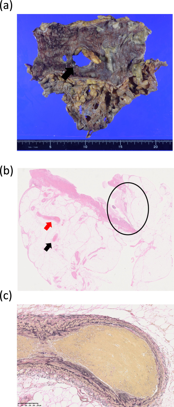
Fig. 4.

Pathological findings of the resected specimen. A total of 17 cm of the transverse colon was resected, and 2 perforation sites of 25 and 7 mm in diameter were identified (arrow). The mucosa around the perforation sites was necrotic (a). The area enclosed in the circle, shows the perforation site. Microcirculatory thrombosis was found in the mesenteric veins (arrow) (b, HE staining, ×3.9). Higher-power field of microcirculatory thrombosis in the mesenteric vein, indicated by a red arrow in b (c, EVG staining, ×77)
