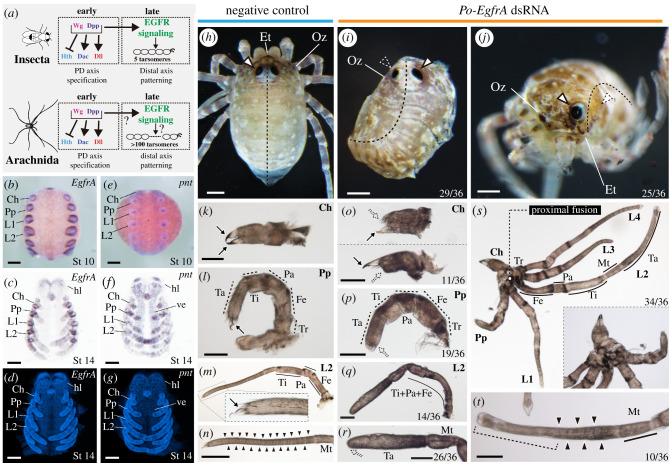
Figure 4.
Po-EgfrA knockdown affects dorsal patterning, eyes and appendage formation. (a) Gene regulatory network specifying PD axis and distal appendage patterning in insects and arachnids. Po-EgfrA (b–d) and Po-pnt (e–g) in situ hybridization wild-type expression. (b,e) Whole-mount stage 10 embryos, merged brightfield and Hoechst nuclear staining, ventral view. (c,f) Flatmounts, stage 14 embryos, brightfield, ventral view. (d,g) Hoechst nuclear counter staining. (h) Negative control hatchling in dorsal view. (h,j) Hatchlings from Po-EgfrA dsRNA-injected treatment (mosaic, left side affected). (b) Hatchling in dorsal view. Note dorsal fusion on the left side of the body (n = 29/36). (c) Hatchling in frontal view, with the left eye absent. A subset of Egfr phenotypes showed eye reduction (25/36) (k–n) Appendage flat mounts of negative control hatchlings, in lateral view. (k) Chelicera. (l) Pedipalp. (m) L2. Inset: detail of the claw. (n) Tarsus of L2. (o–t) Appendage flat mounts of hatchlings of Po-EgfrA dsRNA-injected treatment, in lateral view. (o) Chelicerae with a reduced fixed finger (upper panel), movable finger (lower panel) or both (n = 11/36). (p) Pedipalps lacking claw (n = 19/36). (q) L2, exhibiting podomere fusions proximal to the tarsus (n = 14/36). (r) Distal end of L2, exhibiting claw and tarsomere reduction (n = 26/36). (s) Proximal fusion in adjacent appendages (Ch–L4) (n = 34/36). Inset: Detail of fused coxae. (t) Tarsus of leg 2 shown in (r). Weakly affected legs lacked claws and distal tarsal joints (brackets) but retained proximal joints (n = 10/36). Arrow, claw; outlined white arrowhead, eye; dotted white arrowhead, eye defect; solid black arrowhead, tarsomere joints; Ch, chelicera; Et, egg tooth; Fe, femur; hl, head lobe; L1–L4, legs 1–4; Mt, metatarsus; Oz, ozophore; Pa, patella; Pp, pedipalp; Ta, tarsus; Ti, tibia; Tr, trochanter; ve, ventral ectoderm. Scale bars: 100 µm. (Online version in colour.)

