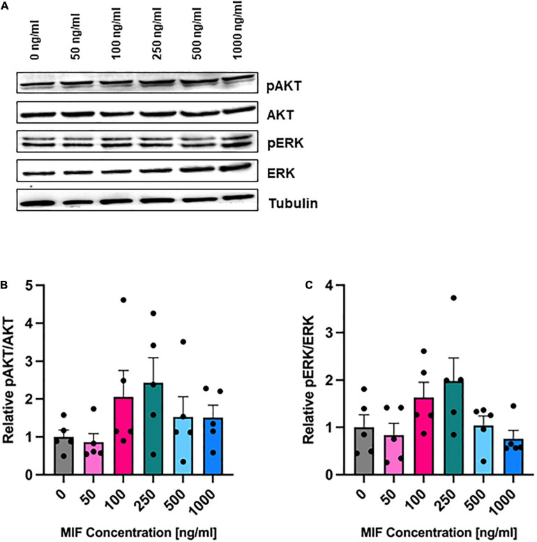
FIGURE 6.
AKT and ERK phosphorylation after MIF stimulation. ASCs were cultured in growth medium at normoxic oxygen levels of 21% O2 for 24 h and then stimulated with various concentrations of exogenous MIF for 15 min. The control (0 ng/ml) did not receive any exogenous MIF. Cell lysates were obtained and Western blot analysis was performed and evaluated using the AIDA software. Tubulin was detected for total protein standardization. (A) Blots are representative of all Western blots performed to detect levels of AKT and ERK activation. Analyses were performed on five biological replicates (n = 5). Graphs display (B) relative pAKT/AKT and (C) relative pERK/ERK normalized to the activation level of unstimulated cells set to one. Data are mean values ± SEM (one-way ANOVA, Holm-Šídák’s multiple comparisons test, not significant).

