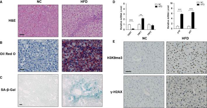
FIGURE 1.

The senescence of hepatocyte occurred in the liver of hamsters with non‐alcoholic fatty liver disease. Male LVG Golden Syrian hamsters were randomly separated into following two groups: group 1, NC group (Standard diet); group 2, HFD group. Liver sections were stained with H&E (A), Oil Red O (B) and SA‐β‐gal (C). Representative photographs are shown. Scale bar, 50 μm (A,B), 100 μm (C). (D) qRT‐PCR analyses of mRNA expression of TERT, TRF1, TRF2, p16 and p21 respectively. Data are represented as mean ±SEM. Significance: ∗∗ P <.01 versus group 1, ∗∗∗ P <.001 versus group 1. (E) Protein expression of H3K9me3 and γ‐H2AX in vivo was investigated by immunohistochemistry on paraffin sections of hamster livers. Scale bar, 50 μm
