FIGURE 3.

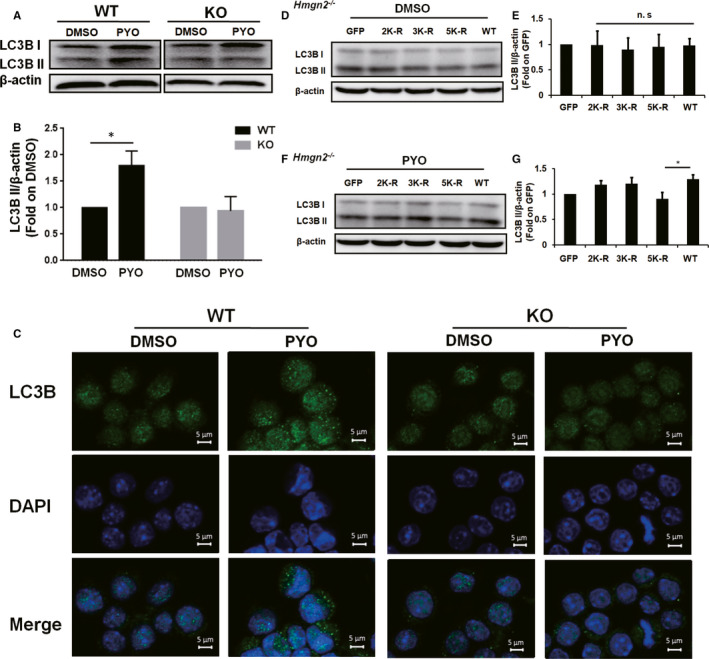
Effect of HMGN2ac on the PYO‐mediated autophagy in the RAW 264.7 cells. The WT and KO RAW 264.7 cells were treated with 50 μM PYO or DMSO for 6 h. (A) Western blot showing LC3B II protein in the RAW 264.7 cells with or without Hmgn2 −/− upon PYO (50 μM, 6 h) or DMSO treatment, and (B) densitometric analysis showing relative expression normalized to that of the DMSO group. (C) Confocal microscopy images displaying the amount of intracellular LC3B puncta (green fluorescence, 630x) with or without Hmgn2 −/− upon PYO (50 μM, 6 h) or DMSO treatment. The nucleus was stained by DAPI (blue fluorescence, 630x), scale bar = 5 μm. (D, F) KO RAW 264.7 cells were transfected, respectively, with the GFP, 2K‐R, 3K‐R, 5K‐R and WT HMGN2 plasmids using jetPRIME for 24 h and then incubated both with 50 μM PYO and with DMSO for 6 h. The Western blot analysis showing the LC3B II protein. (E, G) Densitometric analysis showing relative expression normalized to that of the GFP plasmid. Data are expressed as mean ± SD, *P < 0.05, and n.s indicates no statistical difference, n = 3
