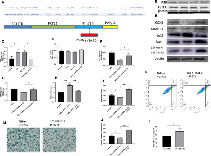
FIGURE 4.

MiR27a inhibits apoptosis, the inflammatory response and matrix degradation by targeting FSTL1. (A) The target genes of miR27a were predicted by miRWalk. (B, C, D) NP cells were transfected with miR27a mimics or NC for 24 h and then treated with 10 ng/mL TNF‐α. After 24 h, FSTL1 (B, C) and P38 (B, D) protein expression was detected by Western blotting. (E, F, G, H, I, J) NP cells were transfected with miR27a mimics for 24 h and then treated with 10 ng/mL TNF‐α alone or in combination with 100 ng/mL rhFSTL1. After 24 h, MMP13 (E, G), Bcl2 (E, H), Bax (E, I), caspase 3 (E, J) and COX2 (E, F) protein expression was detected by Western blotting. (K, L) NP cells were transfected with miR27a mimics for 24 h and then treated with 10 ng/mL TNF‐α alone or in combination with 100 ng/mL rhFSTL1. After 24 h, apoptosis was detected by flow cytometry. (M) NP cells were transfected with miR27a mimics for 24 h and then treated with 10 ng/mL TNF‐α alone or in combination with 100 ng/mL rhFSTL1. After 3 d, extracellular matrix deposition was detected by Alcian blue staining. Statistical analysis was based on at least three different biological samples and three technical replicates; P < .05 was considered to be statistically significant. Representative images are shown
