
FIGURE 5.
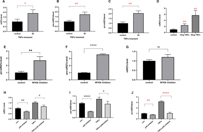
TNF‐α up‐regulates miR27a expression through the P38 pathway. (A, B, C) NP cells were treated with TNF‐α for 8 h, and miR27a (A), pre‐miR27a (B) and pri‐miR27a (C) were measured by qPCR. (D) NP cells were treated with different concentrations of TNF‐α, and miR27a expression was measured by qPCR after 8 h. (E, F, G) NP cells were treated with an inhibitor of NF‐κB (5 µmol/L) for 8 h, and pri‐miR27a (E), pre‐miR27a (F) and miR27a (G) expression was measured by qPCR. (H&I&J) NP cells were treated with an inhibitor of P38 at 10 µmol/L for an hour followed by treatment with TNF‐α for 8 h. Then, miR27a (H), pre‐miR27a (I) and pri‐miR27a (J) expression was measured by qPCR. Statistical analysis was based on at least three different biological samples and three technical replicates; P < .05 was considered to be statistically significant. Representative images are shown