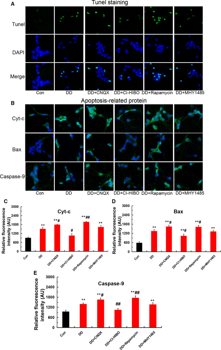
FIGURE 5.

Mitophagy‐induced hippocampal neurons apoptosis under the simulated DD conditions. (A) Hippocampal neurons apoptosis detected by Tunel staining in each group(magnification:200×). (B‐E) Apoptotic proteins of Cyt‐c, Bax and caspase‐9 detected by immunofluorescence staining in each group (magnification:200×; n = 3; **P <.001 vs Con; # P < .05, ## P < .01 vs DD)
