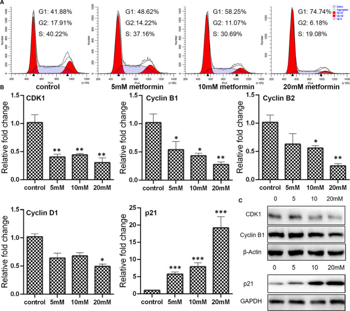
FIGURE 2.

Determination of cell cycle progress in metformin‐treated cells. (A) Cell cycle distribution in breast cancer cells treated with metformin for 24 h was measured by propidium iodide (PI) staining using flow cytometry. (B) Expression of cyclins and cyclin‐dependent kinases as determined by qPCR (mean ± SEM of duplicate experiments). *P < .05 vs control, **P < .01 vs control and ***P < .001 vs control. (C) Expression of CDK1, cyclin B1 and p21 as evaluated by Western blot analysis. Grayscale analysis for Western blot could be seen in Figure S4
