Fig. 5.

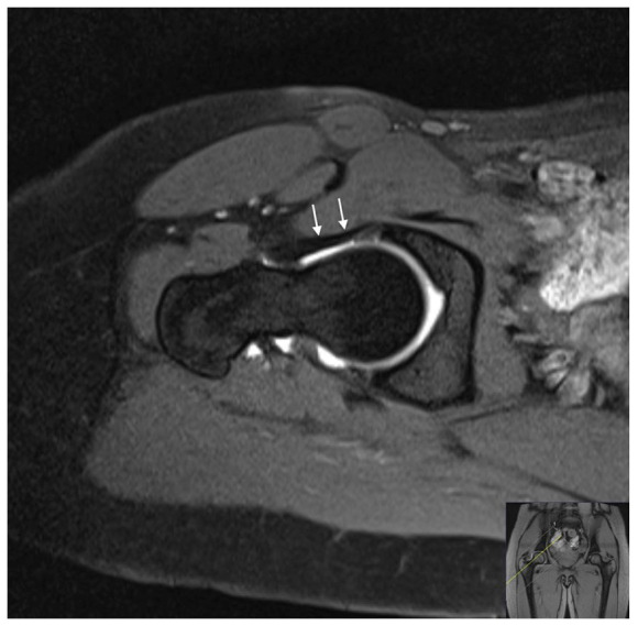
MRI arthrography. Oblique axial T1-weighted fat-suppressed image showing the iliofemoral ligament (white arrows) as a thick band lying anteriorly to the capsule.
Note. MRI, magnetic resonance imaging.

MRI arthrography. Oblique axial T1-weighted fat-suppressed image showing the iliofemoral ligament (white arrows) as a thick band lying anteriorly to the capsule.
Note. MRI, magnetic resonance imaging.