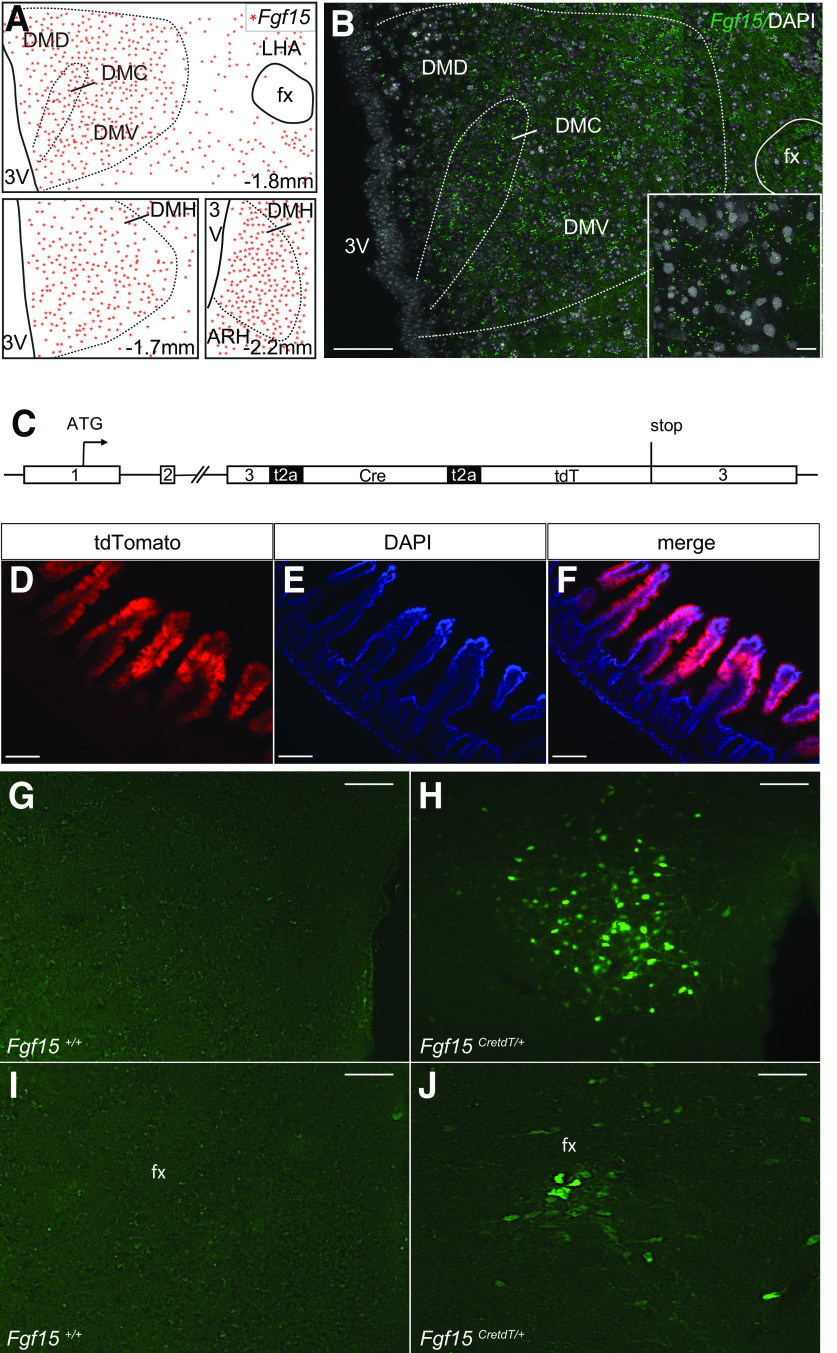
Figure 1.
Reporter mice to characterize Fgf15 neurons. A: Camera lucida cartography of Fgf15 expression in the DMH at the indicated bregma (mm). DMC, DMH compact part; DMD, DMH dorsal part; DMV, DMH ventral part; fx, fornix; LHA, lateral hypothalamic area; 3V, third ventricle. B: In situ hybridization fluorescence detection of Fgf15 mRNA in the dorsal, compact, and ventral divisions of the DMH at bregma −1.8 mm. Scale bar = 50 μm. Inset: Scale bar = 25 μm. C: Structure of the modified Fgf15 allele for the monocistronic expression of Fgf15, Cre, and tdTomato. D–F: Immunofluorescence detection of tdTomato (A), DAPI (B), and merge signals (C) in the ileal villi of Fgf15CreTdT/+ mice. Scale bar = 100 μm. G–J: Immunofluorescence detection of eYFP in the DMH (B and C) and PeF (D and E) of Fgf15+/+ and Fgf15CretdT/+ mice injected with recombinant AAV2-EF1a-DIO-EYFP. Scale bar = 50 μm.

