Figure 4.
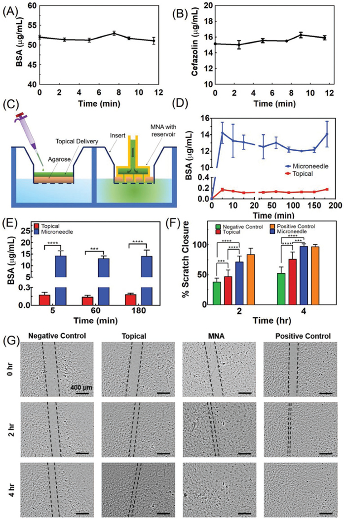
Characterizing the drug release and its effect on cellular cultures. A,B) The concentration of BSA and cefazolin in solutions perfused through the engineered bandage and MNAs over time. The results suggest an insignificant change in concentration (n = 3 for each solution). C) Schematic of the two-compartment in vitro model used for simulating chronic wounds covered by a crust and necrotic tissue used for comparing the topical and MNA-based drug delivery. D) The cumulative concentration of the BSA in the bottom chamber representing the wound bed after the administration of 20 μg mL−1 solution of BSA through 2 mm thick agarose gel (3% w/v) within a cell culture insert (n = 3 for each group). E) The cumulative drug concentration after 5, 60, and 180 min postdrug administration (***P < 0.001, ****P < 0.0001). F,G) Scratch assay on the culture of HUVECs receiving the following treatments: 1) 50 ng mL−1 of VEGF in the culture medium (positive control), 2) no VEGF (negative control), 3) equivalent to 50 ng mL−1 delivered topically, and 4) equivalent to 50 ng mL−1 delivered using the MNAs (n = 5 for each group) (***P < 0.001, ****P < 0.0001). Representative micrographs are shown in (G).

