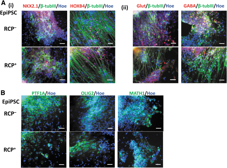
FIG. 2.
Characterization of cerebellar spheroids differentiated from an episomal iPSC line (EpiPSC). With (RCP+) or without (RCP–) indicates the presence of RA, CHIR, and PMR. The representative fluorescent images of (A) common neural markers: (i) NKX2.1 (ventral) and HOXB4 (hindbrain) costained with β-tubulin III (β-tubIII); (ii) GLUT and GABA costained with β-tubulin III (β-tubIII); (B) the cerebellar markers PTF1A, OLIG2, and MATH1. All the images were taken using the Olympus IX70 fluorescent microscope. Scale bar: 100 μm. iPSC, induced pluripotent stem cell; PMR, purmorphamine; RA, retinoic acid.

