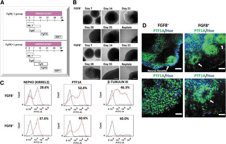
FIG. 5.
The effects of FGF8 on the differentiation of cerebellar spheroids using iPSK3 cells. (A) FGF8 was added during weeks 2 and 3 of the differentiation. (B) The phase contrast images of the cerebellar spheroids for 35 days were taken using the Olympus IX70. Scale bar: 200 μm. (C) Representative flow cytometry histograms of cerebellar molecular layer markers NEPH3 and PTF1A. Black line: negative control, red line: marker of interest. (D) The confocal fluorescent images of the cerebellar spheroids for PTF1A were taken using the Zeiss LSM 880 microscope. The white arrows point toward the neural rosettes. Scale bar: 100 μm (tops) and 50 μm (bottoms).

