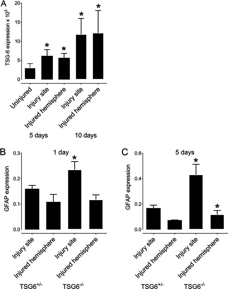
Fig. 1.
TSG-6 and GFAP expression after PBI. TSG-6 and GFAP mRNA expressions were quantified in the injury site and the injured hemisphere after PBI. A TSG+/− mice were subjected to PBI, and the injury site and remaining injured hemisphere were collected 5 and 10 days after injury for analysis of TSG-6 expression. B, C TSG+/− and TSG-6−/− mice were subjected to PBI, and the injury site and remaining injured hemisphere were collected 1 day (B) and 5 days (C) after injury for analysis of GFAP expression. * = p ≤ 0.05 comparing TSG-6−/+ and TSG-6−/− mice

