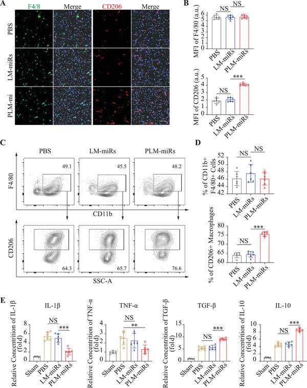
Figure 6.

PLM‐miRs promote reparative polarization of macrophages in vivo. A) CLSM images of MI/R injured heart sections showing the total (F4/80+) and M2 subtype (CD206+) macrophages after treated by PBS, LM‐miRs, or PLM‐miRs. Scalar bar, 50 µm. B) MFI quantification of F4/80 (total) and CD206 (M2) in (A) (n = 6). C) Flow cytometry assay and D) statistical analysis of M2 subtype (CD206+) macrophages isolated from MI/R induced murine hearts after treated by PBS, LM‐miRs, or PLM‐miRs (n = 6). E) ELISA analysis of IL‐1β, TNF‐α, TGF‐β, and IL‐10 concentrations in heart homogenate of MI/R induced mice after treated by PBS, LM‐miRs, or PLM‐miRs (n = 6). Results are presented as mean ± SD. NS P > 0.05, *P < 0.05, **P < 0.01, ***P < 0.001.
