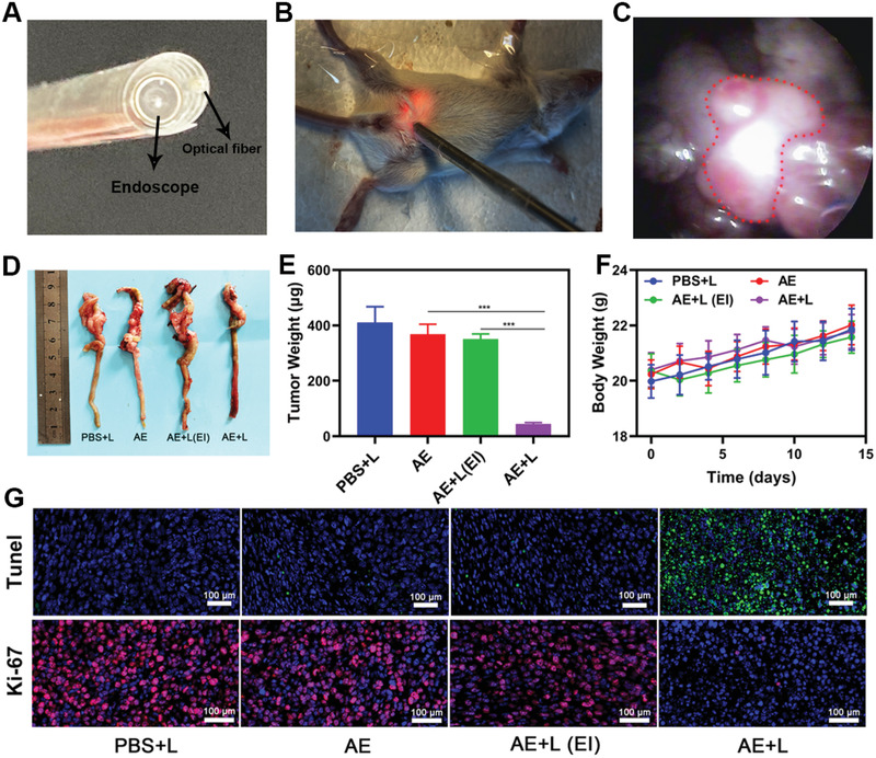
Figure 4.

A) Development of an interventional device composed of an endoscope and an optical fiber. B) Images of mice bearing orthotopic CT26 tumors undergoing interventional PDT. C) Orthotopic tumor position within the abdominal cavity as visualized via endoscopy. The tumor is marked using a red dashed line. D) Images of resected orthotopic CT26 tumors in the indicated treatment groups. E) Tumor volume and F) body weight curves following the indicated treatments in mice bearing orthotopic CT26 tumors. G) Representative TUNEL and Ki‐67 stained tumor sections from mice in the indicated treatment groups. *P<0.05, **P<0.01, ***P<0.005; Student's t‐test.
