
Figure 2.
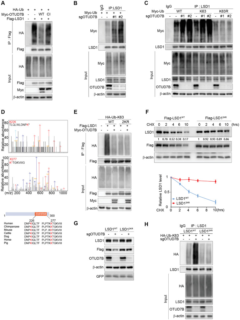
OTUD7B is a bona fide LSD1 deubiquitinase. A) IB analysis of WCL and Flag immunoprecipitate derived from HEK293T cells transfected with the indicated constructs and treated with BTZ for 5 h before harvesting. B) HEK293T cells stably expressing OTUD7B sgRNAs were transfected with Myc‐tagged Ub, followed by IP and IB analysis as indicated. C) HEK293T cells stably expressing OTUD7B sgRNAs were co‐transfected with the indicated Ub constructs, followed by IP and IB analysis as indicated. D) HEK293T cells were co‐transfected with Flag‐tagged LSD1, HA‐tagged ubiquitin, and Myc‐tagged OTUD7B. Cell lysates were subjected to IP and mass spectrometry analysis to identify ubiquitination sites. The recovered LSD1 peptide and the ubiquitination sites were highlighted in red (upper panel). Lower panel: A schematic diagram showing two evolutionarily conserved lysine residues (K226 and K277) within the SWIRM domain of LSD1. E) HEK293T cells were co‐transfected with the indicated plasmids, followed by IP and IB analysis as indicated. F) LSD1WT‐ or LSD12KR‐reconstituted MDA‐MB‐231 cells were treated with CHX for the indicated times. Lysates were subjected to IB analysis as indicated (top panel). The graph shows the quantification of protein levels (bottom panel). G) IB analysis of WCL derived from the indicated LSD1 reconstituting cells expressing OTUD7B sgRNA lentiviruses. H) LSD1‐reconstituted cells stably expressing the indicated sgRNAs were transfected with K63‐only Ub plasmid, followed by IP and IB analysis as indicated. Data were presented as mean ± SEM of three independent experiments.