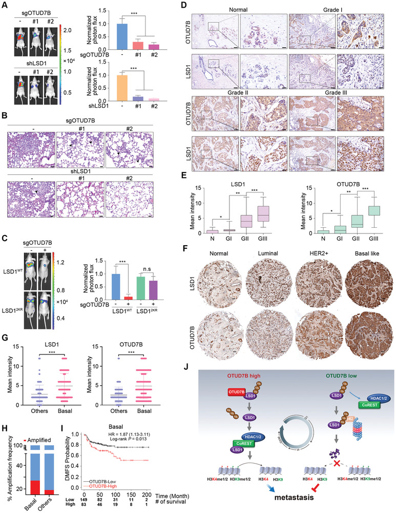
Figure 7.

OTUD7B promotes metastasis via deubiquitinating LSD1. A) Parental or C) LSD1 reconstituting LM2 cells infected with the indicated lentiviruses were injected into the mammary fat pads of nude mice. Representative bioluminescent images of mice with spontaneous lung metastasis (left panel), quantification (right panel), and (B) representative H&E staining analysis of lung metastasis is shown. Results represent mean ± SEM. n = 8 mice per group. Scale bars: 40 µm. ***P < 0.001. P values were calculated by two‐tailed unpaired t test. D) Representative IHC staining of LSD1 and OTUD7B in normal breast tissues (n = 26) and breast carcinomas (n = 379) (histological I, II, and III). Scale bars: 100 µm for low magnification (10×) and 25 µm for high magnification (40×). E) Quantification of LSD1 and OTUD7B intensity in (D). P values were calculated by two‐tailed unpaired t test. * P < 0.05, ** P < 0.01, and *** P < 0.001. F) Representative IHC staining of LSD1 and OTUD7B in normal tissue (n = 22) and the indicated breast carcinoma subtypes (luminal = 76, HER2+ = 49, and basal‐like = 130). Scale bars: 100 µm. G) Quantification of OTUD7B and LSD1 intensity in (F). *** P < 0.001. P values were calculated by two‐tailed unpaired t test. H) TCGA DNA sequencing results showing that the OTUD7B gene is amplified at higher frequencies in basal‐like subtype (27%) compared to other subtypes (19%). I) Kaplan–Meier plots analysis of distant metastasis free survival rates (DMFS) in basal‐like breast cancer patient with high or low OTUD7B mRNA levels. Patient number at risk at different times of analysis is shown at the bottom of the plots. J) The working model of OTUD7B‐mediated LSD1 deubiquitination in coordinating LSD1 turnover, LSD1/CoREST corepressor complex assembly, and the subsequent impact on cell cycle and cancer metastasis.
