Figure 3.

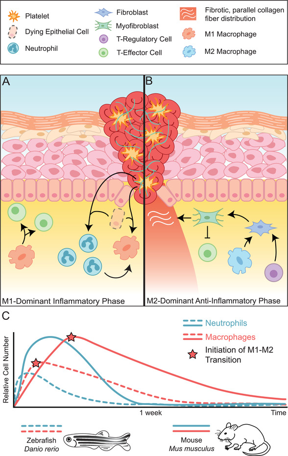
The phases of the inflammatory response to wound healing and differences observed in regenerative zebrafish versus non‐regenerative mouse. A) The proinflammatory response to wound healing is dominated by M1 macrophage polarization. In this phase, inflammatory M1 macrophages (orange), T‐effector cells (green), and neutrophils (teal) are recruited to the wound site. Signals released from dying epithelial cells and platelets drive this recruitment, encouraging neutrophils and monocytes to migrate into the area. M1 macrophages differentiate from monocytes, their polarization mediated by neutrophil signals. Macrophages then secrete cytokines that recruit T‐effector cells. B) An M2‐dominant phase begins when macrophages take on M2 polarization phenotypes (light blue) facilitated by T‐regulatory cells (purple). T‐regulatory cells and M2 macrophages secrete factors such as TGF‐β which leads to the differentiation of fibroblasts into myofibroblasts and the secretion of ECM and suppression of inflammatory T‐effector cells. C) The inflammatory response is markedly different in regenerative systems as observed in the zebrafish, D. rerio (dashed lines) than in the mouse M. musculus (solid lines). Comparatively, numbers of neutrophils (blue) and macrophages (red) are lower in zebrafish and peaks in relative cell number take place at earlier time points. The initiation of the transition from M1 to M2 polarization phenotypes in macrophages also take place earlier on (red stars).
