
Figure 5.
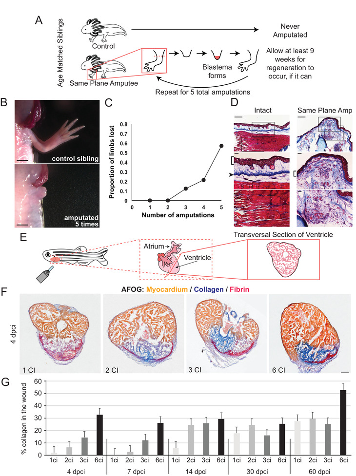
Regenerative decline and fibrosis after repeat injury in axolotl and zebrafish. A) Experimental overview – age matched siblings were used for repeat amputation experiments whereby control animals were never amputated and experimental animals underwent amputation of both forelimbs. The limbs were allowed at least 9 weeks to regenerate, if they were able, and were then challenged to a repeat amputation in the same amputation plane. This process was repeated until the animal was subject to 5 total rounds of amputation. B) Representative bright‐field photos of the control sibling limb (left) and a limb that failed to regenerate in an experimental animal after repeat amputation (right). C) Cumulative distribution plot of loss of the ability to regenerate beyond the plane of amputation. D) Limb stumps that fail to regenerate exhibit persistent collagen deposition, as observed with Masson's trichrome stain. Intact specimen with no amputations (left) shows normal collagen distribution. Failed regenerates following a repeat same plane amputation (right) showed extensive scar tissue, as evidenced by collagen deposition proximal to the plane of amputation. Middle and lower panels are higher magnification views of the images in the top panels. Brackets indicate epidermis, arrowhead indicates dermis in the control. Scale bars in the top panels are 500 µm and scale bars in the middle and bottom panels are 100 µm. B‐D) Adapted with permission.[ 153 ] Copyright 2017, The Authors, published by Nature Portfolio. E) Representative schematic of cryoinjury in zebrafish, D. rerio. F) Representative transversal sections of regenerating, cryoinjured zebrafish hearts at 4 days post‐cryoinjury stained with acid Fuchsin and Orange‐G (AFOG) reagent showing myocardium (beige), fibrin (red), and collagen (blue). Multiple cryoinjuries show more collagen deposition. Scale bar = 100 µm. G) Histogram depicting the percentage of collagen observed in the wound area after cryoinjury. Early in regeneration, the amount of collagen deposition is progressive with increasing numbers of cryoinjury exposure. N ≥ 4 hearts, 3 sections per heart. F,G) Adapted with permission.[ 154 ] Copyright 2020, The Authors, published by Springer Nature.