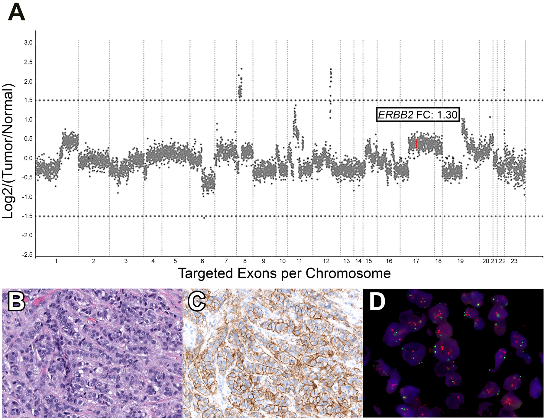
Figure 1.

Representative 2018 ASCO/CAP ISH Group 4 case. A. Copy number plot determined by Memorial Sloan Kettering-Integrated Mutation Profiling of Actionable Cancer Targets (MSK-IMPACT) next-generation sequencing assay, with each dot representing a probe set, and y-axis values showing the normalized log2 transformed fold change (FC) of tumor versus normal. In this Group 4 case, ERBB2 (red dots) FC is 1.30, indicating no amplification. B. Hematoxylin and eosin-stained section displaying invasive carcinoma of no special type. C. HER2 immunohistochemical-stained slide showing equivocal results (score, 2+) with complete membranous staining of weak to moderate intensity D. HER2 fluorescence in situ hybridization (red signal, HER2; green signal, CEP17), demonstrating a HER2/CEP17 ratio of 1.6 and average HER2 copy number of 4.2, classified as 2018 ASCO/CAP ISH Group 4.
