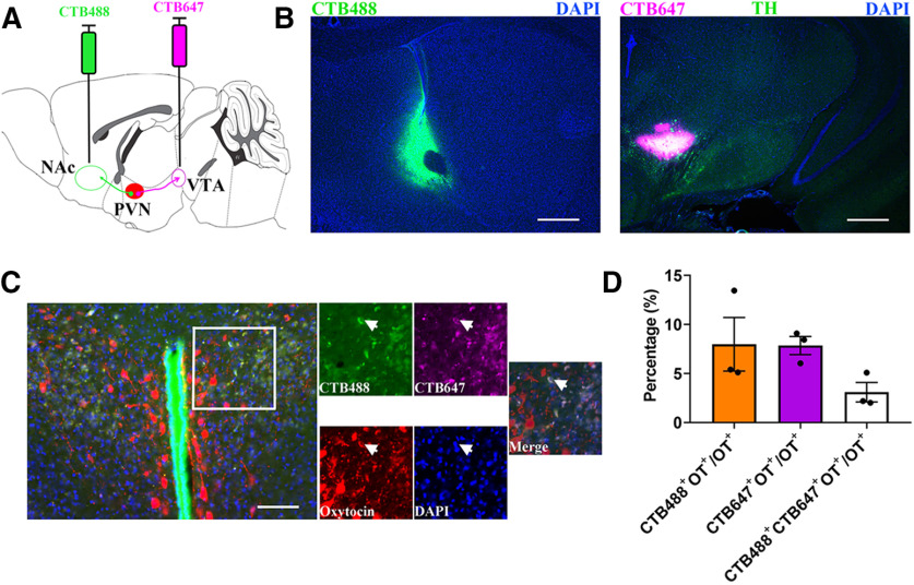
Figure 1.
Anatomically distinct OT neurons project from PVN to the NAc and VTA. A, Experimental schematics for determine collaterals between pairwise injected PVN OT projections. B, Histology of CTB488 injection sites in the NAc (left) and CTB647 injection sites in the VTA (right). Scale bar: 500 μm. TH, tyrosine hydroxylase; DAPI, 4',6-diamidine-2'-phenylindole dihydrochloride. C, An example with three-labeled PVN OT cells (red) is shown after injection of CTB-488 (green) into NAc and CTB-647 (violet) into VTA (indicated by arrowheads). Scale bar: 100 μm. D, Quantification of CTB + and overlapping neurons in all PVN OT population; n = 3.

