
Figure 1.
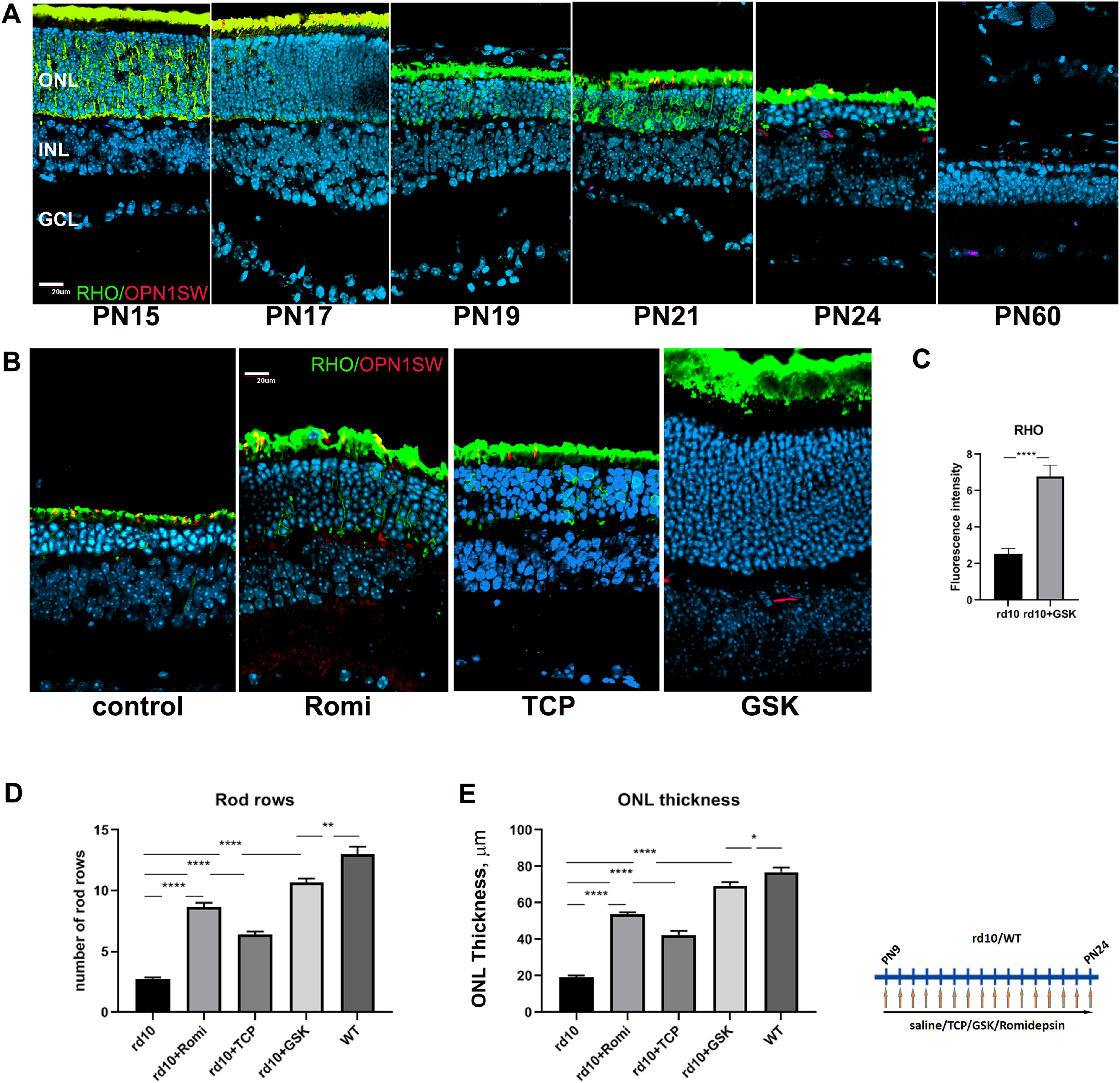
Treatment of rd10 mice with inhibitors specific for LSD1 and HDAC1 leads to neuroprotection and preservation of rod photoreceptors. A, Immunofluorescence microscopic images of sections of retinas from rd10 mice from P15 to P60 stained with RHO (green), OPN1SW (red), and nuclear counterstained with Hoechst33358 (blue); GCL, Ganglion cell layer. Scale bar, 20 μm. B, Immunofluorescence microscopic images of retina sections from P24 rd10 mice treated from P9 until P24 with inhibitors for HDAC1 (romidepsin) or LSD1 (TCP and GSK) or only saline (control), stained with RHO (green), OPN1SW (red), and nuclear counterstained with Hoechst33358. C, Image quantification of immunofluorescence intensity for RHO was conducted for four biological and three technical replicates (±SEM) for the rd10 retinas treated with GSK or saline (control); ****p < 0.0001. D, Rods rows were counted in central retina for P24 mice treated from P9 until P24 with inhibitors for LSD1 (TCP and GSK) and HDAC1 (romidepsin) or only with saline (WT and rd10) for three to five biological and three technical replicas (±SEM); **p < 0.01, ****p < 0.0001. E, ONL thickness was measured in the central retina for P24 mice treated from P9 until P24 with inhibitors for HDAC1 (romidepsin), LSD1 (TCP and GSK), or only with saline (WT and rd10) for three to five biological and three technical replicates (±SEM) for each sample; *p < 0.05, **p < 0.01, ***p < 0.001, ****p < 0.0001.