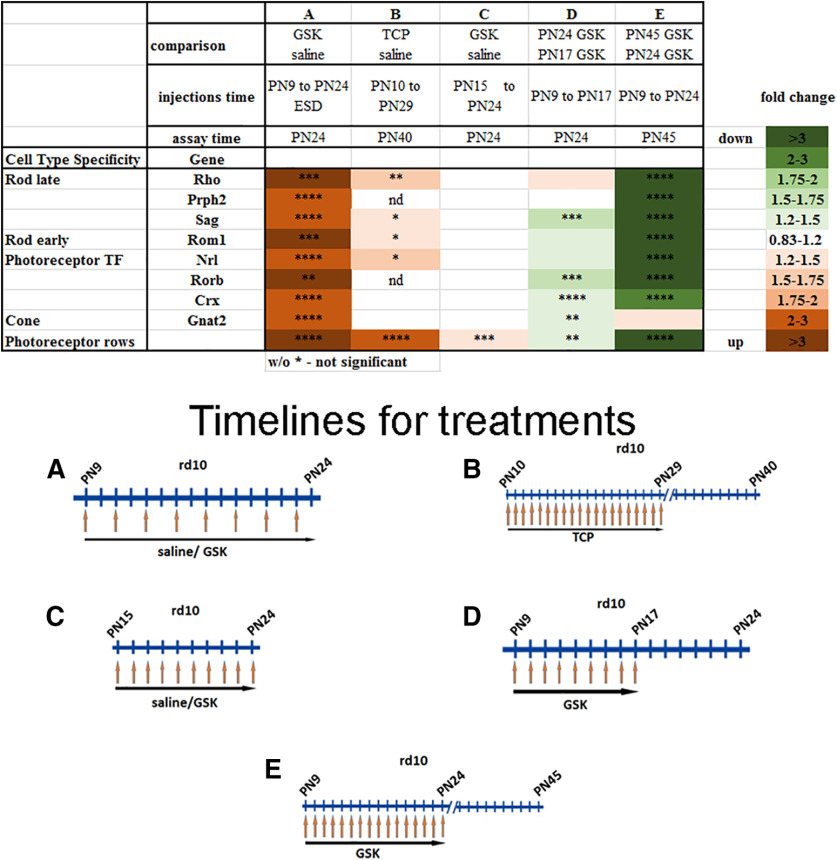
Figure 6.
Changes in expression of retina gene markers are close correlates with rod preservation if alternative time windows were used for i.p. injection of epigenetic inhibitors. Heatmap of expression of different groups of genes measured by RT-PCR and rod rows counted in central retina for retinas from mice treated with inhibitors for LSD1 (TCP and GSK) for three to five biological and three technical replicas (±SEM). **p < 0.01, ****p < 0.0001. The relative expression level for each gene was calculated by the 2-ΔΔCt method and normalized to GAPDH. *p < 0.05, **p < 0.01, ***p < 0.001, ****p < 0.0001 with fold increase in orange or decrease in green. Column A, rd10 mice were treated with GSK from P9 until P24 each second d, assayed at P24, and compared with controls treated with saline only. Column B, rd10 mice were treated with TCP from P10 until P29, assayed at P40, and compared with controls treated with saline only. Column C, rd10 mice were treated with GSK from P15 until P24, assayed at P24, and compared with controls treated with saline only. Column D, rd10 mice litter was treated with GSK from P9 until P17; half litter assayed at P24, half assayed at P17, and compared P24 to P17. Column E, rd10 mice litter was treated with GSK from P9 until P24: half litter assayed at P24, half assayed at P45, and compared P45 to P24.

