Figure 4.

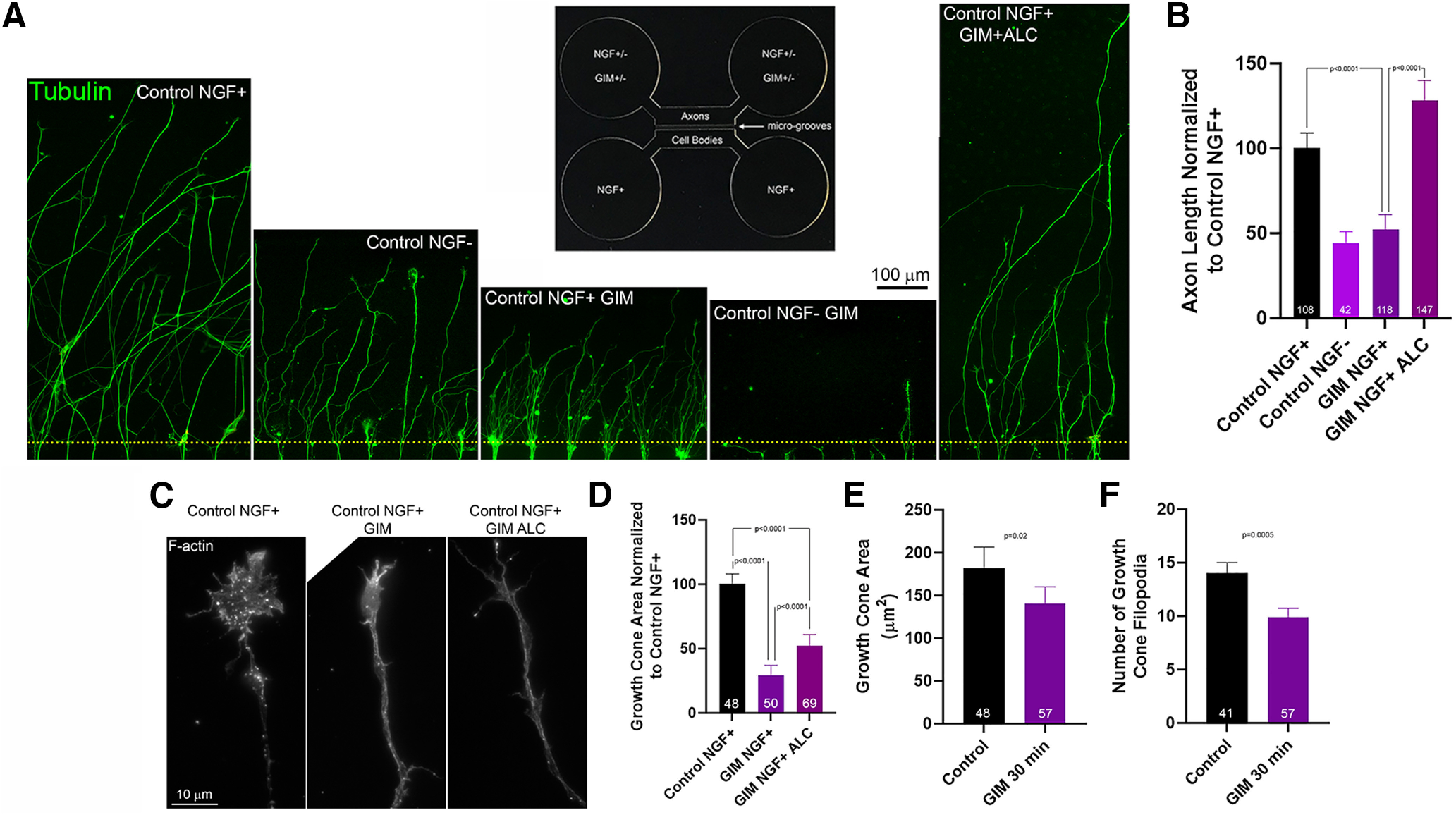
Inhibition of glycolysis in distal axons impairs axon extension in the presence of NGF and blocks it in the absence of NGF. A, Representative examples of axon extension to the axon chamber of microfluidic chambers. Inset, Design of the microfluidic chambers. Yellow dots represent the entry points into the axon chambers. B, Measurements of axon length in axon chambers. Bonferroni multiple comparisons tests. C, Examples of actin filaments (F-actin) in growth cones and distal axons in axon chambers. D, Growth cone measurement area in axon chambers as in A. Bonferroni multiple comparisons tests. Effect of a 30 min treatment with GIM or control medium in the distal chambers after axons had already grown into the chamber on growth cone area (E) and number of filopodia (F). Welch t tests. Mean ± SEM is shown throughout.
