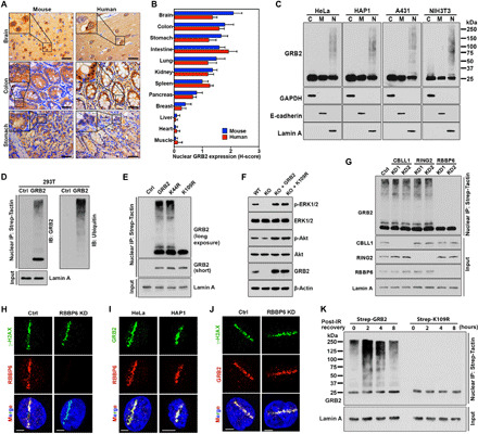
Fig. 1. nGRB2 is poly-ubiquitinated by RBBP6 at the K109 site.

(A) IHC analysis of GRB2 expression in mouse and human tissue samples. Scale bars, 50 μm. (B) H-score quantification of nGRB2. (C) GRB2 expression patterns by subcellular fractionations. Cytoplasm (C), plasma membrane (M), and nucleus (N). (D) Strep-GRB2 with Strep-tag alone (Ctrl) was precipitated from the nuclear extracts of human embryonic kidney (HEK) 293T cells and analyzed with GRB2 or ubiquitin antibodies. (E) Strep-tagged K44R or K109R mutant GRB2 was precipitated from the nuclear extracts of HEK293T cells and immunoblotted with anti-GRB2 antibody. (F) Wild-type (WT), GRB2-KO cells, and KO cells reconstituted with WTGRB2 or K109RGRB2 were immunoblotted with indicated antibodies. (G) Strep-GRB2 was precipitated from the nuclear extracts of CBLL1-, RING2-, or RBBP6-KD HEK293T cells and immunoblotted with indicated antibodies. (H) Micro-irradiated RBBP6-KD HeLa cells were analyzed with the indicated antibodies. (I) Micro-irradiated cells analyzed with indicated antibodies. (J) As (H), except with indicated antibodies. (K) HEK293T cells stably expressing either WT or K109R mutant GRB2 were exposed to 5-Gy IR (152 s) and then either immediately lysed (t = 0). Following indicated recovery time, precipitated GRB2 from nuclear extracts was immunoblotted with anti-GRB2 antibody. All data are representative of three independent experiments. Scale bars, 5 μm.
