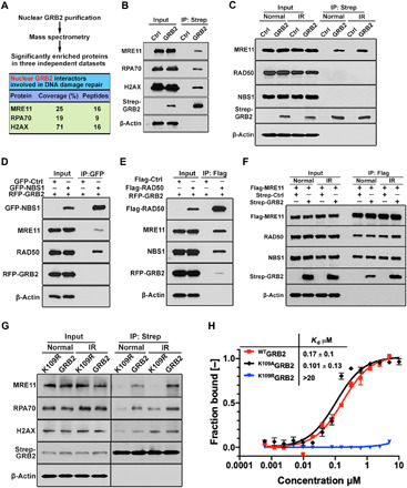
Fig. 2. Interactions of nGRB2 with DNA damage repair factors.

(A) MS identifies DDR protein associated with GRB2. Mean of coverage and unique peptides from three independent data sets are shown. (B) Strep-GRB2 precipitated from HEK293T cells followed by immunoblotting with indicated antibodies. (C) Strep-GRB2 expressed HEK293T cells treated with or without 5-Gy IR were lysed immediately followed by Strep-Tactin precipitation and immunoblotting with indicated antibodies. (D) HEK293T cells were cotransfected with red fluorescent protein (RFP)–GRB2 and green fluorescent protein (GFP)–NBS1 or GFP alone as control, precipitated with GFP-trap beads, and immunoblotted with indicated antibodies. (E) HEK293T cells expressing Flag-RAD50 or Flag-Ctrl were cotransfected with RFP-GRB2 for 24 hours. Unperturbed cells were immunoprecipitated with Flag-M2 beads and immunoblotted with indicated antibodies. (F) HEK293T cells expressing Flag-MRE11 were cotransfected with Strep-GRB2 or Strep-Ctrl for 24 hours. Unperturbed or IR-treated (5 Gy) cells were immunoprecipitated with Flag-M2 beads and immunoblotted with indicated antibodies. (G) Strep-Tactin precipitation of WT and K109R mutant Strep-GRB2 from HEK293T cells that were unperturbed or IR treated (5 Gy) and lysed, followed by Western blot detection with the indicated antibodies. (H) MST isotherms of 100 nM Atto488–labeled WT, K109R, or K109A mutant to titrating concentrations of human MRE11 (residues 1 to 411). All data are representative of three independent experiments.
