
Fig. 5. pH2AX creates a platform for GM complex recruitment.
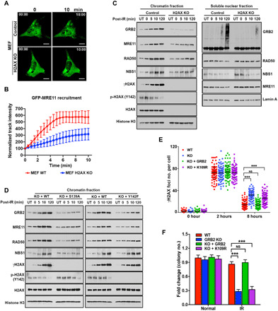
(A) Representative images of live-cell GFP-MRE11 recruitment in WT and H2AX-KO MEFs. Scale bars, 10 μm. (B) GFP-MRE11 track intensity values from 20 cells at every 30 s were quantified, and normalized average intensities are shown with SDs. (C) WT and H2AX-KO MEFs were either untreated (UT) or irradiated with 5-Gy IR (152 s) and then lysed immediately (0 min) or allowed to recover for the indicated time. Chromatin and soluble nuclear fractions were analyzed by Western blotting with indicated antibodies. (D) H2AX-KO MEF cells reconstituted with either WT-H2AX or S139A and Y142F mutant were treated as above, and the chromatin fractions were analyzed by Western blotting with the indicated antibodies. (E) Quantitative analysis of γH2AX foci from GRB2-KO HeLa and KO cells stably reconstituted with WT or K109R mutant GRB2, treated with 5-Gy IR, and then fixed at the indicated times. The numbers of γH2AX foci were counted and represented. (F) Colony survival assay of WT HeLa cells, GRB2-KO cells, and stable GRB2-KO cells reconstituted with WT or K109R mutant, cultured for 10 days after exposure to IR (1 Gy). The significance was analyzed by two-sided Student’s t test. ***P ≤ 0.001 and ****P ≤ 0.0001; NS, not significant.