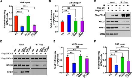
Fig. 6. GRB2 KO suppresses HDR.

(A) DR-GFP reporter assay for control (WT), GRB2-KO (KO), and indicated reconstituted U2OS cells. The percentage of GFP-positive cells was determined by fluorescence-activated cell sorting (FACS) 72 hours after transfection. Normalized data are shown. (B) Normalized data showing a measurable increase in Alt-EJ in GRB2-KO cells. The up-regulation was suppressed with either WT GRB2 or K109R mutant reconstitution. (C) HEK293T cells overexpressing Flag-tagged POLQ was immunoprecipitated immediately after 5-Gy IR treatment or without treatment and immunoblotted with indicated antibodies. (D) Parental control (WT), GRB2-KO (KO), WTGRB2 (KO + GRB2), and K109RGRB2 (KO + K109R) reconstituted HEK293T cells were transfected with Flag-tagged XRCC1. After 24 hours, XRCC1 was immunoprecipitated and the resulting coimmunoprecipitants were analyzed by Western blotting with indicated antibodies. β-Actin was used as a loading control. (E) Normalized data showing a significant reduction in NHEJ in GRB2-KO cells that was rescued by reconstitution of either WT GRB2 or K109R mutant. (F) Normalized data showing GRB2-KO and reconstituted cells for single-stranded annealing (SSA) repair. The K109R mutant only partially rescued the GRB2-KO phenotype. The significance was analyzed by two-sided Student’s t test. *P ≤ 0.05, **P ≤ 0.01, ***P ≤ 0.001, and ****P ≤ 0.0001; NS, not significant.
