
Figure 2.
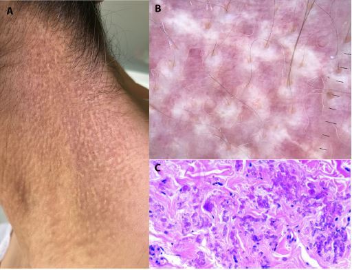
Dermatologic, dermoscopic, and histopathologic examination. (A) Dermatological examination showed confluent yellowish papules on the neck. (B) Dermoscopic examination revealed reticulated yellow-to-white clods on a light red background, along with disfocused linear irregular vessels. (C) Histopathological examination showing dermal accumulation of swollen clumped fibers.