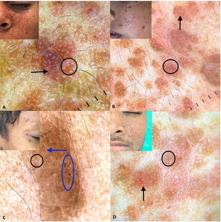
Figure 1.

(A–D) Dermoscopy showing yellow-white dots (black circle) over a brown to reddish-brown background (black arrow) with blurred vessels. At places there are unevenly distributed specks/dots of brown pigmentation (blue arrow). Few lesions show surface crypts (blue oval). Inset displays clinical images of the represented dermoscopic findings. (Dermlite©, 3Gen Inc., San Juan Capistrano, CA, U.S.A., DL200 hybrid, Polarized mode, magnification 10x). Images were captured with Dermlite adaptor for Iphone©11.
