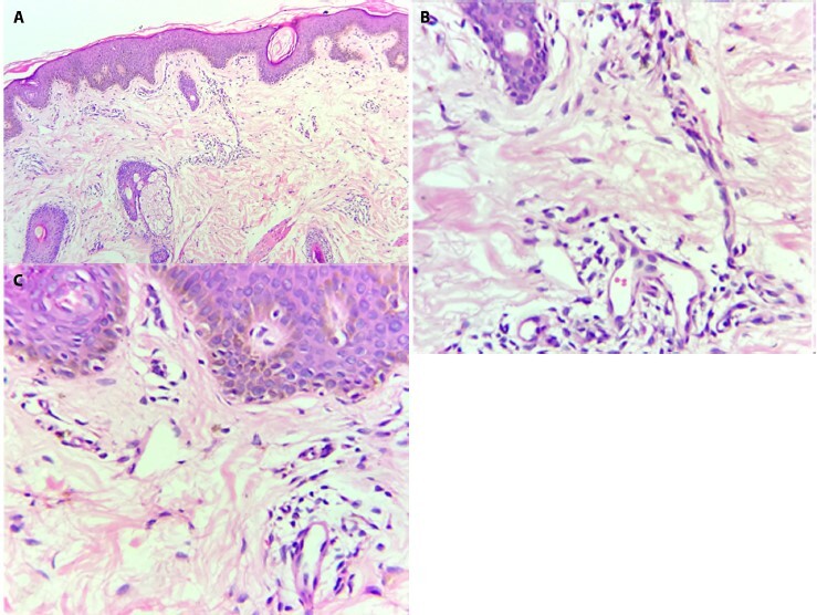
Figure 2.

(A) Epidermis showing follicular plugging, patchy to linear melanocytic hyperplasia. Dermis showing increased number of blood vessels surrounded by mild lymphocytic infiltrate. Increased collagen fibers in dermis (H&E, × 10). (B) Increased dermal melanophages (H&E, × 40). (C) Linear melanocytic hyperplasia (H&E, × 40).
