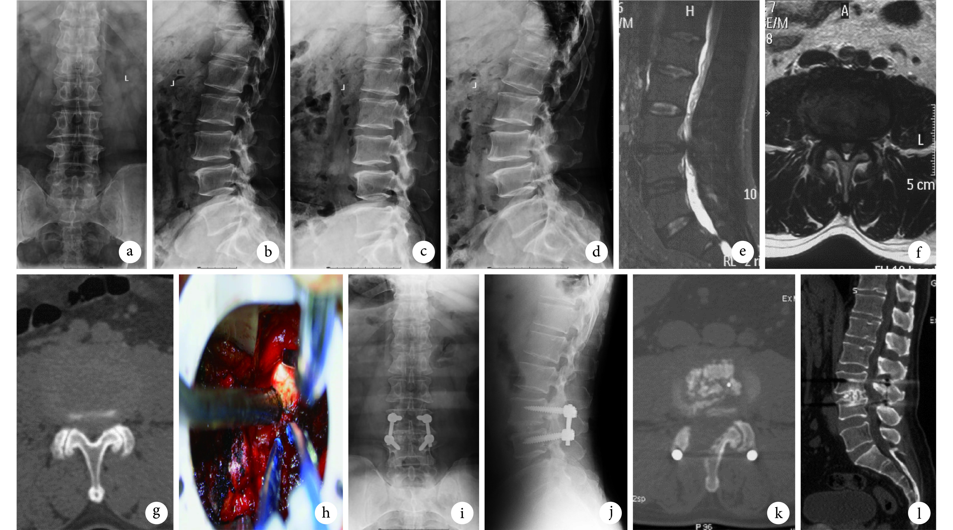
图 2.
A 55-year-old male patient with lumbar spinal stenosis at L3, 4
U 组患者,男,55岁,L3、4 腰椎管狭窄症
a、b. 术前腰椎正侧位 X 线片;c、d. 术前腰椎动力位 X 线片示 L3、4 失稳;e、f. 术前腰椎 MRI 示 L3、4 椎管狭窄;g. 术前 CT 示中央管、双侧侧隐窝狭窄;h. 术中通道内操作减压;i、j. 术后 1 年腰椎正侧位 X 线片;k、l. 术后 1 年 CT 示椎管容积明显增大,L3、4 椎间骨性融合
a, b. Anteroposterior and lateral X-ray films of lumbar spine before operation; c, d. Flexion and extension X-ray films showed the instability of L3, 4; e, f. MRI showed the spinal stenosis at L3, 4 before operation; g. CT showed the central canal and bilateral recess stenosis before operation; h. Decompression under tube; i, j. Anteroposterior and lateral X-ray films at 1 year after operation; k, l. CT showed that the canal volume significantly increased and the interbody fusion at L3, 4 obtained at 1 year after operation

