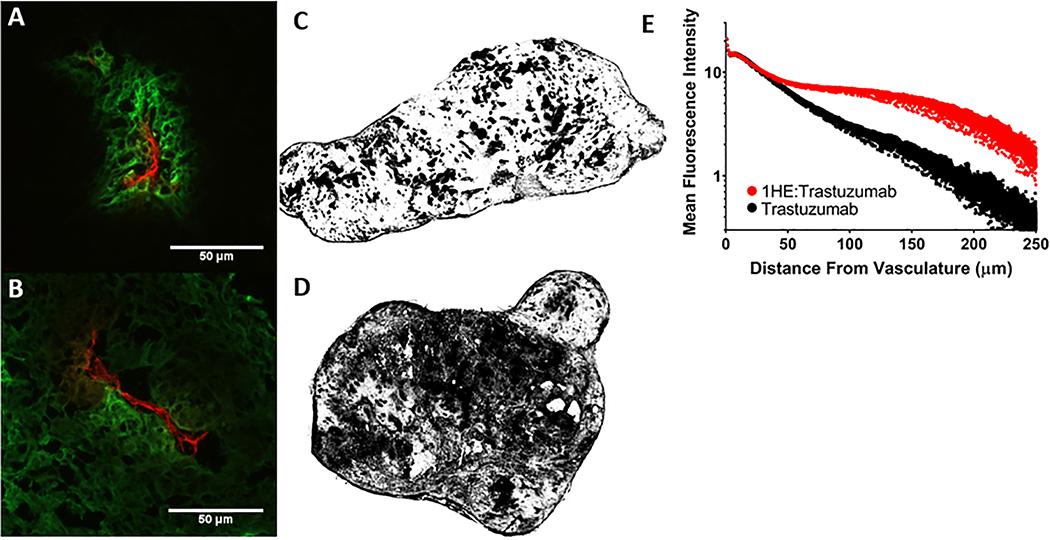
Figure 4: Impact of 1HE co-administration on trastuzumab distribution in SK-OV3 xenografts.
(A) Trastuzumab administered alone (green) is restricted around vasculature (red) whereas (B) 1HE co-administration increased trastuzumab tumor penetration as indicated by the diffuse staining from the point of extravasation. Whole tumor sections of trastuzumab and trastuzumab:1HE are shown in C and D respectively, images were converted to black and white with regions of trastuzumab positive fluorescent staining two-fold greater than background appearing in black. In comparison to tumors treated with trastuzumab alone. 1HE co-administration dramatically increased the fraction of the tumor that stained positive for trastuzumab. Trastuzumab associated fluorescence staining (MFI) as a function of distance from the nearest blood vessel is shown with and without 1HE co-administration. Individual points represent the mean of all pixels at a given distance from the vasculature. Similar fluorescence intensity is observed for trastuzumab administered alone and with 1HE up to 30 μm from the vasculature. Starting at 30 μm the trastuzumab:1HE group has greater staining intensity which extends up to 250 μm from the vasculature.

