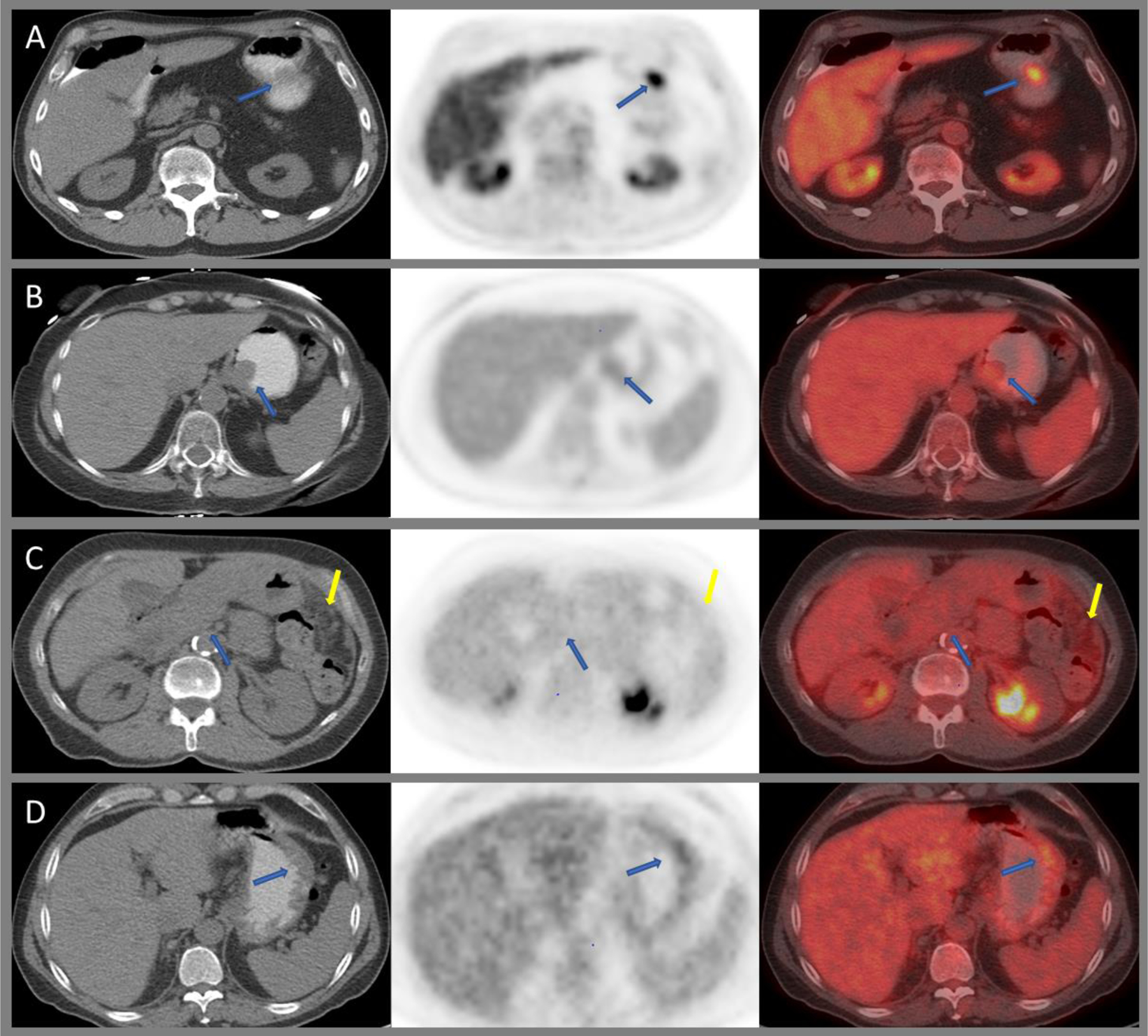
Figure 4:

Axial CT, PET, and fused images of 18F-FDG PET/CT demonstrating intensely avid gastric schwannoma along the greater curvature of the stomach (A), mildly avid leiomyoma at the gastroesophageal junction (B), diffuse low-grade uptake within an infiltrating poorly differentiated adenocarcinoma with signet ring cell features (blue arrows) and peritoneal carcinomatosis (yellow arrows), (C) and mild diffuse uptake within a biopsy proven gastric MALT lymphoma (D).
