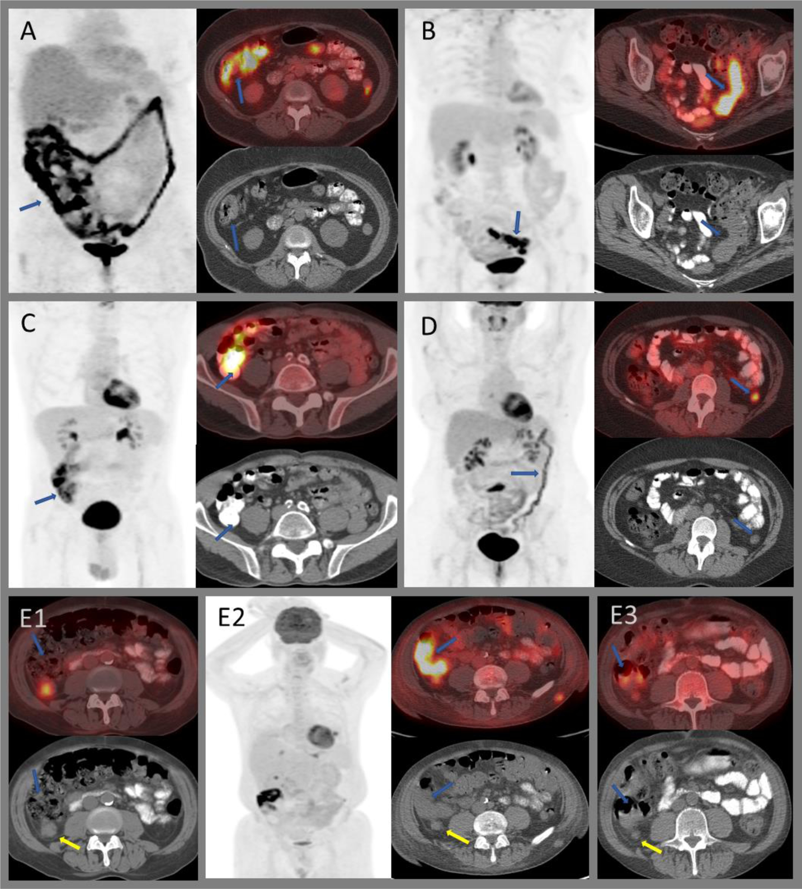
Figure 7:

Variable FDG uptake within the colon. A) Intense diffuse uptake throughout the large bowel in patient on metformin. B) Segmental intense uptake within the sigmoid colon with bowel wall thickening in a patient with known ulcerative colitis without active symptoms. C) Intense uptake secondary to attenuation artifact from oral contrast media. D) Moderately intense diffuse uptake within the distal large bowel in a patient with active ulcerative colitis. E) Post-radiation colitis of the hepatic flexure (E2) in a patient who underwent radiation therapy for a moderately intense right posterior peritoneal metastasis (E1; yellow arrows). Post-radiation, the nodule decreased in size; however, intense uptake is seen within the adjacent hepatic flexure (E2; blue arrows). Bowel uptake resolved on subsequent imaging with residual low-grade uptake within the peritoneal nodule (E3).
