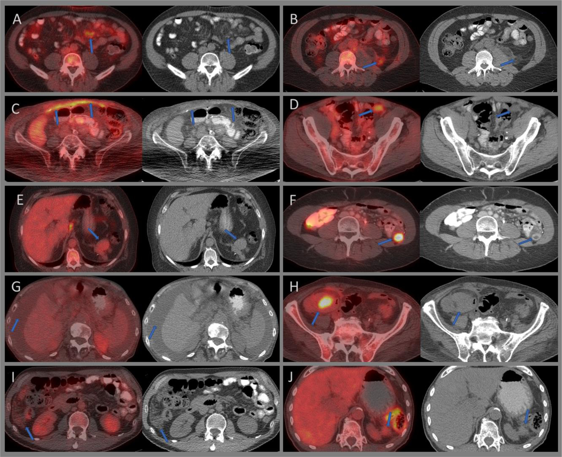
Figure 9:

Axial fused and CT images of 18F-FDG PET/CT demonstrating moderately intense mesenteric panniculitis (A), left lower quadrant fat necrosis (B), intense uptake relating to surgical mesh hernia repair (C), moderately intense uptake with a hernia plug (D), mildly avid splenules at the pancreatic tail (E), intensely avid transposed ovary within the left paracolic gutter (F), low-grade malignant ascites from unknown primary (G), intensely avid post-surgical abscess adjacent to the ascending colon (H), minimally avid peritoneal mesothelioma within the right lilac fossa (I), and a minimally avid recurrent peritoneal metastatic deposit at the splenectomy bed from a pancreatic tumor (J).
