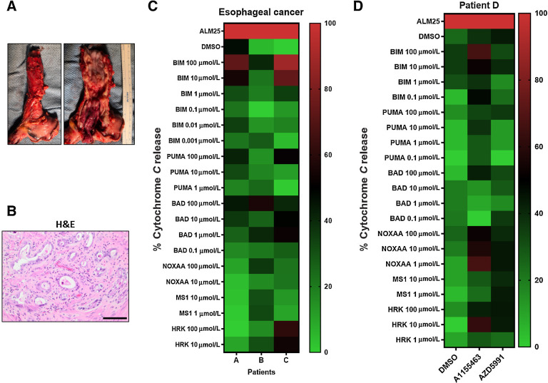
Figure 5.
BH3 profiling in patients with EAC. A, An EAC of the distal esophagus after surgery. Left, The gross appearance of the intact esophagus. Right, The mucosal surface with the tumor. B, H&E staining of the resected specimen shows atypical cytologic features including increased nuclear/cytoplasmic ratio, pleomorphism, prominent nucleoli, and intraluminal necrotic debris (scale bar = 200 μmol/L). C, Heatmap showing BH3 profilings in three patients with EAC. D, DBP with 0.2 μmol/L A1155463 and AZD5991 for 20 hours ex vivo from a metastatic pleural implant that was not radiated with the primary esophageal tumor.

